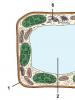
Plant tissue diagram. Plant tissues. Types of plant tissue. II. Learning new material
Plant tissue
Namefabrics
Structure
Location
Functions
Educational tissue (meristem)
The meristem is formed by living, small, tightly closed cells, with a large nucleus, dense cytoplasm and small vacuoles.
Participates in the formation of new cells and the differentiation of these cells into cells of other tissues.
apical
Cone of growth in the buds, seed embryo, at the tips of the roots
1. Ensures organ growth in length.
2. Thanks to cell division and differentiation, tissues of roots, shoots, leaves, and flowers are formed.
Lateral (cambium)
Located between the wood and the phloem of the stems and roots
Thickening of the stem and root.
cover tissue
Located on
surface of the whole plant
1. Protects the plant from drying out and other unfavorable
influences.
2. Participates in the breathing process.
3. Participates in the exchange of substances between the environment.
Peel (epidermis)
It consists of a layer of living, tightly packed cells with a thickened wall, without chloroplasts. The skin of leaves and green shoots contains stomata.
Located on
leaf surfaces,
young shoots, all parts of the flower
1. Protection of organs from drying out and microorganisms.
2. Stomata provide gas and water exchange in plants.
Cork
Made up of dead cells
Covers the stems
perennial plants,
rhizomes, tubers
1. Protection against shocks
temperatures, mechanical
influences, pests.
2. Multi-layer cork
forms on the surface
stem protective cover containing lentils
for gas and water exchange.
Crust
Complex of multilayer cork and other dead tissues that replaces the epidermis in perennial plants
Covers the lower part of the trunks, well defined
at the bark oak
Mechanical protection
damage, changes
temperatures, pests,
microorganisms.
The main tissue is parenchyma
The main tissue usually consists of living, thin-walled cells that form the basis of organs
1. Photosynthesis.
2. Supply of nutrients.
Assimilation
textile
Leaf tissue contains chloroplasts
Mainly in green
leaves and young shoots
1. Photosynthesis
2. Gas exchange
Storage
Consists of homogeneous
thin-walled cells in which proteins, fats, carbohydrates and other reserve substances are deposited in the form of starch grains and oil droplets. They often have large vacuoles containing cell sap.
It is found in the stems of woody plants
(core), root vegetables,
tubers, bulbs, fruits and seeds
1. Accumulation of spares
nutrients.
2. Cells of primary tissues are capable of turning into secondary tissue
fabric, due to which
vegetative occurs
plant propagation.
Conductive fabric
Consists of elongated cells
It is a component of wood (xylem) and bast
(phloem)
Provides transport
nutrients from
root to leaves (upward current), from leaves to root (downward)
Xylem (wood)
Xylem containsvessels (dead elongated cells, devoid of transverse partitions, the walls of which are impregnated with lignin, which gives
vessels additional hardness)
Located in the wood of the stem, the conducting zone
root, leaf veins
The main conducting tissue of higher vascular plants. It is also involved in the transport of minerals (upstream), the storage of nutrients and performs a support function
Phloem (bast)
Consists of sieve tubes with satellite cells. Sieve
the tubes are formed by living cells, the transverse partitions of which are pierced with small holes that form a “sieve”. Cells do not have nuclei, but they have cytoplasm. Satellite cells are connected to sieve tubes and perform a trophic function (nutrition, enzyme synthesis)
Forms conductive
bunches in the phloem along the stem, root, leaf veins
Conducts dissolved
organic matter,
formed in leaves
(downward current), into the stem, root, flowers, fruits
Mechanical
textile
Mechanical tissue cells have thick, thickened and lignified membranes that fit tightly together
Mechanical tissues are mainly located in the stem; in the root, they are present only in the center. Surrounds vascular bundles
Gives strength to plant organs, counteracts
rupture or fracture, form a framework that supports the plant organs
excretory
textile
Consists of cells that form
and secreting various substances (secrets)
Secret allocation
Ferrous
hairs
Living cells forming
long outgrowths - hairs filled with liquid secretion
On the surface of the leaves
stems (stinging
nettle cells, glandular hairs of geranium). At the base of the petals
1. Release of substances,
protecting against being eaten by animals, microorganisms, evaporation
2. Release of odorous substances that attract pollinating insects
Sunbirds
Living cells filled with sweet, often strong-smelling contents
Flower (most often at the base of the petals)
Secretion of nectar that attracts
pollinating insects
Resin and milky passages
Dead elongated cells filled with resin or milky juice
Coniferous wood, dandelion stem
Protection against microorganisms,
damage, eating
animals
Main fabrics constitute the bulk of the plant's body. They consist of living, relatively few specialized cells, often of a parenchymal form, which is why they are often called parenchymal tissues, or parenchyma. Depending on the function performed, several types of basic tissues are distinguished.
Assimilation tissue (chlorophyll-bearing parenchyma, chlorenchyma) performs the function of photosynthesis. It is located mainly in the leaves and stems of herbaceous plants just behind the epidermis. The cells are living, thin-walled, often parenchymal in shape. 70-80% of the protoplast volume is made up of chloroplasts. Characterized by the presence of intercellular spaces that facilitate gas exchange ( rice. 3.2).
Rice. 3.2. Cross section of a belladonna leaf: 1 – cells of assimilative tissue; 2 – cells filled with crystalline calcium oxalate sand.
Storage parenchyma serves as a site for the deposition of nutrients (starch, proteins, fatty oils). Reserve nutrients can be deposited in living cells of any tissue, but this function is especially pronounced in specialized storage tissues, well developed in seeds, roots, and underground shoots ( rice. 3.3.A). Storage tissues consist of living thin-walled cells, often of parenchymal form.
A type of storage tissue is aquifer parenchyma, which performs the function of storing water. It consists of large living thin-walled cells, usually of parenchymal form. Water is stored in vacuoles due to the high content of mucus, which has a high water-holding capacity. Aquiferous parenchyma is found in the stems and leaves of succulents (cacti, agaves, aloe), in many salt marsh plants (soleros, anabasis, saxaul), and in the leaves of many cereals. A lot of water is contained in the storage tissues of bulbs and tubers.
Airborne parenchyma (aerenchyma) performs the function of ventilation, supplying tissues and organs with oxygen. It is well developed in the submerged organs of aquatic and marsh plants (water lily, egg capsule, calamus, watch). Aerenchyma consists of living cells of various shapes and large intercellular spaces ( rice. 3.3.B).

Rice. 3.3. Storage parenchyma of potato tuber (A) and aerenchyma of the pondweed stem (B): 1 – intercellular space.
Mechanical parenchyma occupies an intermediate position between the main and mechanical tissues. These are living parenchyma cells with a slightly thickened lignified cell wall.
Non-specialized parenchyma (basic parenchyma, non-specific parenchyma) It is a living parenchymal tissue without a pronounced function. This tissue is always present in the body of the plant, making up its largest part.
3.4. Integumentary tissues
Integumentary tissues are located on the surface of plant organs at the border with the external environment. They consist of tightly closed cells and protect the internal parts of the plant from adverse external influences, excessive evaporation and drying, sudden changes in temperature, penetration of microorganisms, and serve for gas exchange and transpiration. According to their origin, they are distinguished from various meristems. primary And secondary covering tissues.
TO primary integumentary tissues include: 1) rhizoderm, or epibleme and 2) epidermis.
Rhizoderm (epiblema) – primary single-layer superficial root tissue. Formed from protodermis– the outer layer of cells of the apical meristem of the root. The main function of the rhizoderm is absorption, selective absorption from the soil of water with mineral nutrition elements dissolved in it. Through the rhizoderm, substances are released that act on the substrate and transform it. Rhizoderm cells are thin-walled, with viscous cytoplasm and a large number of mitochondria (mineral ions are actively absorbed, with energy consumption, against the concentration gradient). A characteristic feature of rhizoderm is the formation of some cells root hairs– tubular outgrowths, unlike trichomes, not separated by a wall from the mother cell ( rice. 3.4). Root hairs increase the absorptive surface of the rhizoderm tenfold or more. The hairs are 1-2 (3) mm long. Rhizoderm is often considered as suction textile.
Rice. 3.4. The tip of the root of Ozhika multiflorum: 1 – root hair.
Epidermis- primary integumentary tissue formed from protodermis shoot growth cone. It covers leaves, stems of herbaceous and young shoots of woody plants, flowers, fruits and seeds. The main function of the epidermis is the regulation of gas exchange and transpiration(evaporation of water by living tissues). In addition, the epidermis performs a number of other functions. It prevents pathogens from entering the plant, protects internal tissues from mechanical damage and gives organs strength. Essential oils, water, and salts can be released through the epidermis. The epidermis can function as an absorptive tissue. It takes part in the synthesis of various substances, in the perception of irritations, and in the movement of leaves.
The epidermis is a complex tissue; it consists of morphologically different types of cells: 1) main cells of the epidermis; 2) closing and subsidiary cells of stomata; 3) trichomes.
Basic cells of the epidermis– living cells of tabular shape. The appearance of cells from the surface is different ( rice. 3.5). The cells are tightly closed, intercellular spaces are absent. The side walls (perpendicular to the surface of the organ) are often tortuous, which increases the strength of their adhesion, less often straight. The epidermal cells of the axial organs and leaves of many monocots are strongly elongated along the axis of the organ. 
Rice. 3.5. Leaf epidermis of various plants (surface view): 1 - iris; 2 - corn; 3 – watermelon; 4 - initial letter.
The outer cell walls are usually thicker than the rest. Their inner, more powerful, layer consists of cellulose and pectin substances; the outer layer undergoes cutinization. A continuous layer of cutin is released on top of the outer walls, forming a protective film - cuticle. In addition to cutin, it contains impregnations of wax, which further reduces the permeability of the cuticle to water and gases. Wax can be deposited in crystalline form and on the surface of the cuticle in the form of scales, rods, tubes and other structures visible only with an electron microscope. This bluish, easily erased coating is clearly visible on cabbage leaves, plums, and grapes. The power of the cuticle, the distribution of waxes and cutin in it determine the chemical resistance and permeability of the epidermis to gases and solutions. In dry climates, plants develop thicker cuticles. Plants immersed in water have no cuticle.
Epidermal cells have a living protoplast, usually with a well-developed endoplasmic reticulum and Golgi apparatus. Most plant species contain leucoplasts in the cytoplasm. Rare chloroplasts are found in aquatic plants, ferns, and inhabitants of shady places (hibiscus). The epidermis most often consists of a single layer of cells. Rarely, two- or multi-layered epidermis is found, mainly in tropical plants living in conditions of variable water supply (begonias, peperomia, ficus). The lower layers of the multilayered epidermis function as water-storing tissue. In some plants, the cell walls may be impregnated with silica (horsetails, grasses, sedges) or contain mucus (flax seeds, quince, plantain).
Stomata– formations for the regulation of transpiration and gas exchange. The stomata consists of two closing cells bean-shaped, between which there is stomatal gap, which can expand and contract. Under the gap there is a large intercellular space - substomatal cavity. The epidermal cells adjacent to the guard cells are often different from the rest of the cells, and are then called side effects, or parastomatal cells(rice. 3.6). They are involved in the movement of guard cells.

Rice. 3.6. Diagram of the structure of the stomata.
Guard and subsidiary cells form stomatal apparatus. Depending on the number of side cells and their location relative to the stomatal fissure, several types of stomatal apparatus are distinguished (Fig. 3.7). In pharmacognosy, types of stomatal apparatus are used to diagnose medicinal plant materials.

Rice. 3.7. Types of stomatal apparatus: 1 – anomocytic; 2 – diacite; 3 – paracytic; 4 – anisocytic; 5 – tetracite; 5 – encyclocytic.
Anomocytic the type of stomatal apparatus is common for all groups of plants, with the exception of horsetails. The side cells in this case do not differ from the rest of the epidermal cells. Diacitic the type is characterized by two subsidiary cells, which are located perpendicular to the stomatal fissure. This type is found in some flowering plants, in particular in most Lamiaceae (mint, sage, thyme, oregano) and cloves. At paracytic Typically, two side cells are located parallel to the guard cells and the stomatal fissure. It is found in ferns, horsetails and a number of flowering plants. Anisocytic the type is found only in flowering plants, in particular, it is found in cruciferous plants (shepherd's purse, yellowwort) and nightshades (henbane, datura, belladonna). In this case, the guard cells are surrounded by three side cells, one of which is noticeably larger or smaller than the others. Tetracite The type of stomatal apparatus is characterized mainly by monocots. At encyclocytic In this type, side cells form a narrow ring around guard cells. A similar structure is found in ferns, gymnosperms and some flowering plants.
The mechanism of movement of guard cells is based on the fact that their walls are thickened unevenly, so the shape of the cells changes as their volume changes. A change in the volume of cells of the stomatal apparatus occurs due to changes in osmotic pressure. The increase in pressure occurs due to the active intake of potassium ions from neighboring cells, as well as due to an increase in the concentration of sugars formed during photosynthesis. Due to the influx of water, the volume of the vacuole increases, turgor pressure increases, and the stomatal fissure opens. The outflow of ions occurs passively, water leaves the guard cells, their volume decreases, and the stomatal fissure closes. In most plants, stomata open during daylight hours and close at night. This is due to the fact that photosynthesis occurs only in light, and it requires an influx of carbon dioxide from the atmosphere.
The number and distribution of stomata vary greatly depending on the plant species and environmental conditions. In most plants their number is 100-700 per 1mm2 of leaf surface. With the help of stomata, the epidermis effectively regulates gas exchange and transpiration. If the stomata are completely open, then transpiration proceeds at the same rate as if there were no epidermis at all (according to Dalton’s law, with the same total area of the holes, the higher the number of holes, the higher the evaporation rate). When the stomata are closed, transpiration is sharply reduced and can actually only go through the cuticle.
In many plants, the epidermis forms external single- or multicellular outgrowths of various shapes - trichomes. Trichomes are extremely diverse, while remaining quite stable and typical for certain species, genera and even families. Therefore, the characteristics of trichomes are widely used in plant taxonomy and in pharmacognosy as diagnostic ones.
Trichomes are divided into: 1) coverts and 2) glandular. Ferrous Trichomes form substances that are considered secretions. They will be discussed in the section on excretory tissues.
Coverts trichomes look like simple, branched or stellate hairs, unicellular or multicellular ( rice. 3.8). Covering trichomes can remain alive for a long time, but more often they quickly die and fill with air.
A thick layer of hairs reflects some of the sun's rays and reduces heating, creating a quiet space near the epidermis, which together reduces transpiration. Often the hairs form a cover only where the stomata are located, for example, on the underside of the leaves of coltsfoot and wild rosemary. Hard, prickly hairs protect plants from being eaten by animals, and papillae on the petals attract insects.

Rice. 3.8. Covering trichomes: 1-3 – simple unicellular, 4 – simple multicellular, 5 – branched multicellular, 6 – simple bicornuate, 7,8 – star-shaped (in plan and in cross-section of the leaf).
It is necessary to distinguish from trichomes, which are formed only from epidermal cells emergents, in the formation of which deeper tissues also take part. These include rose, raspberry, and blackberry thorns covering leaf petioles and young shoots.
TO secondary integumentary tissues include: 1) periderm and 2) crust, or rhytide.
Periderm- a complex multilayer integumentary tissue that replaces the primary integumentary tissues - rhizoderm and epidermis. The periderm covers the roots of the secondary structure and the stems of perennial shoots. It can also occur as a result of healing of damaged tissue by the wound meristem.
The periderm consists of three complexes of cells, different in structure and function. These are: 1) phellem, or cork, performing the main protective functions; 2) phellogen, or suberic cambium, due to the work of which the periderm as a whole is formed; 3) phelloderm, or cork parenchyma, which performs the function of feeding phellogen ( rice. 3.9).

Rice. 3.9. The structure of the periderm of the elderberry stem .
Fellema (cork) consists of several layers of tabular cells, located densely, without intercellular spaces. Secondary cell walls are composed of alternating layers of suberin and wax, making them impermeable to water and gases. The cork cells are dead, they do not have a protoplast and are filled with air. Substances that increase the protective properties of the cork can also be deposited in the cell cavity.
Phellogen (cork cambium)– secondary lateral meristem. It is a single layer of meristematic cells that deposit cork cells on the outside and phelloderm cells on the inside of the organ. Phelloderm (cork parenchyma) refers to the main tissues and consists of living parenchyma cells. However, often the phellogen works one-sidedly, depositing only a plug, while the phelloderm remains single-layered ( rice. 3.9).
The main function of cork is to protect against moisture loss. In addition, cork protects the plant from the penetration of pathogenic organisms, and also provides mechanical protection to the trunks and branches of trees, and phellogen heals the damage caused, forming new layers of cork. Since the cork cells are filled with air, the cork case has low thermal conductivity and provides good protection against sudden temperature fluctuations.
In most trees and shrubs, phellogen is formed in annual shoots already in mid-summer. Most often it arises from parenchymal cells lying just below the epidermis ( rice. 3.9). Sometimes phellogen is formed in the deeper layers of the bark (currant, raspberry). Rarely, epidermal cells, dividing, turn into phellogen (willow, quince, oleander).
Gas exchange and transpiration in organs covered with periderm occur through lentils(rice. 3.10). In places where the lenticels are located, the cork layers are torn and alternate with parenchyma cells, loosely connected to each other. Gases circulate through the intercellular spaces of this performing tissue. Phellogen underlies the supporting tissue and, as it dies, is supplemented with new layers. With the onset of the cold season, phellogen is deposited under the performing tissue cap layer, consisting of cork cells. In spring, this layer breaks under the pressure of new cells. In the trailing layers there are small intercellular spaces, so that the living tissues of tree branches, even in winter, are not tightly separated from the environment.

Rice. 3.10. The structure of the elderberry lentil in a cross section.
On young shoots, lentils look like small tubercles. As the branches thicken, their shape changes. In birch they stretch along the circumference of the trunk and form a characteristic pattern of black dashes on a white background. In aspen, the lenticels take the shape of diamonds.
In most woody plants, the smooth periderm is replaced by a fissured one. crust (rhythid). In pine this happens at 8-10 years, in oak - at 25-30 years, in hornbeam - at 50 years. Only some trees (aspen, beech, sycamore, eucalyptus) do not form a crust at all.
The crust arises as a result of the repeated formation of new layers of periderm in increasingly deeper layers of the cortex. Living cells enclosed between these layers die. Thus, the crust consists of alternating layers of cork and other dead bark tissue ( rice. 3.11).

Rice. 3.11. Cross-section of oak bark .
Dead tissues of the crust cannot stretch, following the thickening of the trunk, so cracks appear on the trunk, but do not reach the deep living tissues. The border between the periderm and the bark is externally noticeable by the appearance of these cracks; this border is especially clear in birch, in which the white birch bark (periderm) is replaced by a black cracked bark. The thick crust reliably protects tree trunks from mechanical damage, forest fires, and sudden changes in temperature.
In any living or plant organism, tissue is formed by cells similar in origin and structure. Any tissue is adapted to perform one or several important functions for an animal or plant organism.
Types of tissues in higher plants
The following types of plant tissues are distinguished:
- educational (meristem);
- integumentary;
- mechanical;
- conductive;
- basic;
- excretory.
All these tissues have their own structural features and differ from each other in the functions they perform.

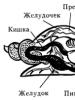
Fig.1 Plant tissue under a microscope
Educational plant tissue
Educational fabric- This is the primary tissue from which all other plant tissues are formed. It consists of special cells capable of multiple divisions. It is these cells that make up the embryo of any plant.
This tissue is retained in the adult plant. It is located:
TOP 4 articleswho are reading along with this
- at the bottom of the root system and at the tops of the stems (ensures plant growth in height and development of the root system) - apical educational tissue;
- inside the stem (ensures the plant grows in width and thickens) - lateral educational tissue;
Plant integumentary tissue
Cover tissue is a protective tissue. It is necessary in order to protect the plant from sudden changes in temperature, from excessive evaporation of water, from microbes, fungi, animals and from all kinds of mechanical damage.
The integumentary tissues of plants are formed by cells, living and dead, that are capable of allowing air to pass through, providing the gas exchange necessary for plant growth.
The structure of plant integumentary tissue is as follows:
- first there is the skin or epidermis, which covers the leaves of the plant, stems and the most vulnerable parts of the flower; skin cells are living, elastic, they protect the plant from excessive moisture loss;
- Next is the cork or periderm, which is also located on the stems and roots of the plant (where the cork layer is formed, the skin dies); The cork protects the plant from adverse environmental influences.
There is also a type of integumentary tissue called crust. This most durable covering tissue, cork, in this case is formed not only on the surface, but also in depth, and its upper layers slowly die off. Essentially, the crust is made up of cork and dead tissue.

Fig. 2 Crust - a type of plant covering tissue
For the plant to breathe, cracks form in the crust, at the bottom of which there are special shoots, lentils, through which gas exchange occurs.
Mechanical plant tissue
Mechanical tissues give the plant the strength it needs. It is thanks to their presence that the plant can withstand strong gusts of wind and do not break under streams of rain or under the weight of fruits.
There are two main types of mechanical fabrics: bast and wood fibers.
Conductive plant tissues
Conductive fabric ensures the transport of water with minerals dissolved in it.
This tissue forms two transport systems:
- upward(from roots to leaves);
- downward(from leaves to all other parts of plants).
The ascending transport system consists of tracheids and vessels (xylem or wood), and vessels are more advanced conductors than tracheids.
In descending systems, the flow of water with photosynthesis products passes through sieve tubes (phloem or phloem).
Xylem and phloem form vascular-fibrous bundles - the “circulatory system” of the plant, which penetrates it completely, connecting it into one whole.
Main fabric
Ground tissue or parenchyma- is the basis of the entire plant. All other types of fabrics are immersed in it. This is living tissue and it performs different functions. It is because of this that its different types are distinguished (information about the structure and functions of different types of basic tissue is presented in the table below).
| Types of main fabric | Where is it located in the plant? | Functions | Structure |
| Assimilation | leaves and other green parts of the plant | promotes the synthesis of organic substances | consists of photosynthetic cells |
| Storage | tubers, fruits, buds, seeds, bulbs, root vegetables | promotes the accumulation of organic substances necessary for plant development | thin-walled cells |
| Aquifer | stem, leaves | promotes water accumulation | loose tissue consisting of thin-walled cells |
| Airborne | stem, leaves, roots | promotes air flow through the plant | thin-walled cells |

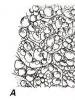
Rice. 3 The main tissue or parenchyma of the plant
Excretory tissues
The name of this fabric indicates exactly what function it plays. These fabrics help saturate the fruits of plants with oils and juices, and also contribute to the release of a special aroma by the leaves, flowers and fruits. Thus, there are two types of this fabric:
- endocrine tissue;
- Exocrine tissue.
What have we learned?
For the biology lesson, 6th grade students need to remember that animals and plants consist of many cells, which, in turn, arranged in an orderly manner, form one or another tissue. We found out what types of tissues exist in plants - educational, integumentary, mechanical, conductive, basic and excretory. Each tissue performs its own strictly defined function, protecting the plant or providing all its parts with access to water or air.
Test on the topic
Evaluation of the report
Average rating: 3.9. Total ratings received: 1552.
Groups of plant cells with a common function, structure and origin are called plant tissues. The most important of them are: integumentary, basic, excretory, conductive, mechanical and educational. Let's consider the structure and functions of plant tissues.
Educational tissues (meristems)
Located in growth zones:
- on the tops of shoots;
- at the tips of the roots;
- along the stems and roots (cambium or lateral meristem, ensures the growth of stems and roots in thickness).
The meristem cells are actively dividing and do not even have time to grow; they are always young, and therefore do not have vacuoles, their walls are thin, and the nucleus is large.
The activity of the apical meristem of bamboo is striking. It grows literally before our eyes, every hour by 2 - 3 cm!
Integumentary tissues
It is known how quickly peeled fruits dry out, or how easily fruits with broken skins become infected with rot. It is the barrier of the integumentary tissue that ensures the safety of the soft parts of the plant.
There are three types of integumentary tissue:
TOP 4 articleswho are reading along with this
- epidermis;
- periderm;
- crust.
Epidermis (skin)- superficial living cells of various organs. Protects underlying tissues and regulates gas exchange and water evaporation by the plant.

Rice. 1. Epidermal cells under a microscope.
Periderm is formed in woody plants when the green color of the shoot turns brown. The periderm consists of cork cells that protect the shoot from frost, microbes and moisture loss.
Crust- dead tissue. It cannot stretch, following the thickening of the trunk, and cracks.
Basic tissues (parenchyma)
There are three types of parenchyma:
- photosynthetic (assimilation);
- aerenchyma, ensures the passage of air into the plant through the intercellular space;
- storing.

Rice. 2. Parenchyma of a green leaf under a microscope.
Conductive fabrics
They ensure the movement of substances in the plant body. The movement is carried out in two main directions:
- rising current , carried out by xylem;
- downward current carried out by phloem.
Xylem and phloem form a continuous, plumbing-like system.

Rice. 3. Scheme of the structure of phloem and xylem.
Phloem vessels are composed of sieve elements, or tubes, - elongated cells, the transverse edges of which are similar to a sieve. The flow of substances passes through the pores of the sieve from one cell to another. The cells in the vessel seem to be placed one on one.
The conducting elements of xylem are also represented by elongated cells, but their pores are also located on the side walls of the cells.
Mechanical fabrics
Provide protection and stability of the plant or its individual parts (fruit seeds). Cell membranes are thickened.
Types of mechanical fabric:
- collenchyma (living cells);
- sclerenchyma (dead cells).
Collenchyma is located in growing leaves and stems; it does not interfere with their growth. Contains elongated cells. After the growth of this part of the plant stops, collenchyma gradually turns into sclerenchyma - it becomes tougher, the shells become lignified and thicken.
Lignification increases the fragility of sclerenchyma. Flax fiber is an exception to the rule; it is not lignified sclerenchyma. That's why flax makes such a soft fabric like cambric.
Excretory tissues
These are tissues that secrete water or some secretion from the plant (essential oil, nectar, resin, salts, etc.). This type of tissue also includes those whose secretions remain inside the plant. These are, for example, lacticifers that contain milky juice in their vacuoles (celandine, dandelion).
Their main function is to remove unnecessary substances and protect. Thus, the resin in coniferous wood protects it from rotting.
Using the table “Plant Tissues” we will briefly summarize what has been said:
|
Fabrics |
Functions |
Features of cell structure |
Location |
|
Integumentary |
Protection and gas exchange |
Tight adhesion of cells to each other |
Plant surface |
|
Educational |
Small, with thin walls |
Apical parts of shoots and roots; |
|
|
Mechanical |
Thickened shells |
Stem, leaf veins |
|
|
Basic |
Photosynthesis, nutrition storage. substances |
Loose arrangement of cells |
The basis of the plant, in all organs; stem center |
|
excretory |
Protection and highlighting |
The structure is varied |
Everywhere |
|
Conductive |
Transport of substances |
Vascular elements |
Everywhere |
What have we learned?
From a 6th grade biology paper, we learned that there are six main types of plant tissues. A plant is a system in which tissues are elements. Each tissue provides some area of plant life. Each tissue is vital; the normal development of the entire plant depends on its successful functioning. Tissue cells are specialized; they have structural features corresponding to the functions they perform.
Test on the topic
Evaluation of the report
Average rating: 4.7. Total ratings received: 548.
Plan
1. Concept of fabric.
2. Plant tissues.
Basic concepts: tissue, differentiation, plant anatomy, forming (meristematic) tissues, integumentary tissues, basic tissues, conductive tissues, mechanical tissues, excretory formations.
Concept of fabric
In most multicellular organisms, during their development, cells begin to differ in structure and functions, that is, they differentiate (from the Latin differentia - difference). Differentiation of cells leads to their specialization to perform certain functions. As a result, tissues are formed.
Textile(from Latin textus, Greek histos) is a system of cells and intercellular substance, united by a common function, structure and origin.
Plant tissue is the object of study of science - plant anatomy (from Greek anatome- dissect).
Plant tissue
A feature of plant tissues, which distinguishes them from animal tissues, is that they have almost no intercellular substance and often contain dead cells. Plant tissues are divided into the following groups:
o forming (meristematic); o basic; o mechanical;
o integumentary; o presenters; o excretory.
Formative, or meristematic(from Greek meristos- dividing), tissues are those tissues that ensure the formation of other tissues and the growth of plants in height and thickness. The cells of this tissue are small and located close to each other. They have thin cell walls and a large nucleus, which ensures cell division.
Formative tissues are located only in certain areas of plants:
o always at the tip of the shoot and the tip of the root - apical meristem, which ensures the growth of these organs in length (Fig. 19, 20);
Rice. 19. Shoot apical meristem:
1 - growth cone;
2 - protodermis;
3 - main meristem;
4 - procambium;
5 - bud rudiment;
6, 7 - leading fabric; 8 - core.
o inside perennial roots and shoots and covers their central part in the form of a cylinder - lateral (lateral) meristem (Fig. 20), which ensures the growth of these organs in thickness;
o at the base of the internodes of the stem of some plants (for example, in cereals) there is an intercalary meristem (Fig. 20), which ensures growth in length due to the elongation of the internodes;
in places where plants are injured - wound meristem, which ensures the regeneration of a particular tissue. The educational activity of the apical meristem is maintained throughout ontogenesis (individual development), so plants are capable of unlimited growth.
There are primary and secondary meristems.
Primary meristems - tissues, as a result of whose activity permanent tissues are formed. The primary meristems include: the procambium, the growth cone of the stem and root, the pericycle and the intercalary meristem.
Secondary meristems - meristems, which are formed from primary meristems or other specialized tissues. The secondary meristems include the fascicular and interfascicular cambium and telogen (cork cambium).

Rice. 20. Scheme of placement of the primary and secondary meristem in dicotyledonous plants:
1 - apical (apical);
2 - lateral (lateral);
3 - plug-in (intercalary);
4 - cambium;
5 - telogen (cork cambium).
Cell division in meristems is regulated by the formation or entry from other tissues of regulatory substances - phytohormones.
Integumentary tissues. The very name of these tissues indicates their location in the plant organism - on the surface of the organs. They separate internal tissues from the external environment and protect them. Depending on the structural features, the integumentary tissues also perform other functions.
The following types of integumentary tissues are distinguished:
o epidermis(from Greek epi - from above, derma- skin), or peel, is a single-layer tissue that covers young plant organs and performs barrier, protective, transpiration (water evaporation), gas exchange, sucking (root hairs) and secretory (hairs, glands) functions.
The insulating properties of the epidermis are enhanced by the formation of a thin waxy film - cuticle (from the Latin cuticula - skin). The cuticle prevents the evaporation of water through the epidermis and ensures that drops of rainwater roll off the leaf.
communication with the environment of the tissues that are located under the epidermis is carried out thanks to the prodykha (Fig. 21), located, for example, in land plants on the underside of the leaf.

Rice. 21. Schematic representation of a respite:
A - open air; B - the outlet is closed.
Stomata are formed by two guard cells capable of closing and opening the gap between them. Unlike other epidermal cells, guard cells contain chloroplasts, which synthesize substances necessary to regulate the opening and closing of stomata. Thanks to the ability of prodichous cells to open and close, the plant regulates the intensity of the processes of transpiration (evaporation of water) and gas exchange.
Epidermal cells often form special outgrowths - hairs. Some of them protect the plant from overheating, others - from herbivorous animals. For example, the stinging hairs of nettle leaves and young shoots produce toxic substances.
A type of epidermis is rhizoderm (from the Greek rhiza - root, derma - skin) - living integumentary tissue, which consists of one layer of living cells with long thin outgrowths - root hairs. It forms the root zone of the plant, through which water and minerals are absorbed from the soil.
Periderm(from the Greek peri - around, derma - skin) - multilayer secondary integumentary tissue of a plant organism. It consists of cork (outer layer), cork cambium (middle layer), felodermis (inner layer).
Traffic jams consists of cells with thickened cell walls in which a fat-like substance - suberin - is deposited, which makes the cell walls impermeable to water and air. This leads to the death of the living contents (protoplast) of the cells. Cork reliably protects the plant during unfavorable periods (for example, winter or during drought).
The connection of lignified stems and roots with the environment is carried out through special holes in the crust - compatriots(Fig. 22). They carry out gas exchange and transpiration. Unlike stomata, stomata are not able to open and close, and on the eve of winter they are clogged with special substances.

Rice. 22. Sochevichka kirkozon (Aristolochia) (for Raven, Zwert, Eichhorn, 1990)
Main fabrics- fabrics located between the covering and leading fabrics. The main tissues consist of living cells with relatively thin cell walls, between which there are usually interstitial spaces. Basic tissues make up the bulk of the body of plants and occupy different positions in them - leaves, bark, pith, etc. Their functional specialization depends on their position in the plant organism. Based on their functions, the main tissues are divided into the following types:
o Assimilation fabric(from the Latin assimilatio - convenient), or guy-renchyma (from the Greek chloros - green, enchyma - tissue), is the main photosynthetic tissue located in the leaves between the upper and lower epidermis (Fig. 23) and young stems in the primary bark . The cells of this tissue contain many chloroplasts (hence the name - renchima guy), in which photosynthesis occurs. This tissue is characterized by a developed system of intercellular air cavities associated with pro-dichos. This ensures gas exchange of tissues involved in photosynthesis.

Rice. 23. Cross section of an oleander leaf (Nerium oleander) (Raven, Evert, Eichhorn, 1990)
Storing fabric- loose tissue, built from living colorless cells with thin cell walls and large vacuoles, in which various compounds necessary for plants accumulate (carbohydrates, proteins, lipids, vitamins, water, organic acids). This tissue is located in the core of the stem, seeds, fruits, modified shoots and roots.
Ventilation fabric or aerenchyma(from Greek polyester- air, enchyma - tissue), - tissue consisting of small cells separated by a well-developed system of large intercliniforms, which are combined into a single ventilation network and promote gas exchange. This tissue is characteristic of plants that live in conditions where there may be a lack of air. Usually these are aquatic and marsh plants, for example, water lilies.
Leading fabrics is a collection of highly specialized cells that are adapted to transport inorganic and organic substances and are the main components of vascular bundles.
The following leading elements are distinguished (Fig. 24):
o Tracheids - these are dead elongated spindle-shaped (prosenchymal) cells with thick, usually lignified walls, with pointed ends, due to which they are connected to each other in longitudinal rows with a large contact area. The cell walls have complex pores (bordered) through which water passes. Tracheids provide an upward flow of water with mineral salts from the underground part of the plant to the above ground. Thanks to the solid cell walls of the trachea, in addition to the leading one, they also provide a supporting function. Tracheids are characteristic of most higher spore plants (except bryophytes) and gymnosperms. In conifers, tracheids are located in the wood mainly in regular radial rows.

Rice. 24. Lead fabric(according to Yakovlev, Chelombitko, 2001): A - tracheids; B 1-5 - different types of vessels; B - sieve-like tubes: 1 - sieve-like cell; 2 - satellite cell; 3 - transverse wall
with pores.
o Vessels - these are dead cells connected in series, the transverse walls between them have disappeared. They provide an upward flow of water with mineral salts from the underground part of the plant to the above ground. The driving forces for the movement of substances are root pressure, transpiration (evaporation of water through stomata), and interaction forces between water dipoles. Thanks to the vital cell walls of the vessel, in addition to the leading one, they also provide a supporting function. The vessels are characteristic of angiosperms and some gymnosperms. The vessels function for several years, after which they are clogged with parenchyma cells and begin to perform a supporting function.
o Sieve-like tubes - these are living (but without a nucleus) elongated cells, sequentially located one above the other in the form of a chain. The transverse walls of these cells have numerous small holes that resemble a sieve (hence their name). Sieve-like tubes “accompany” cells-or-satellites that have nuclei. These cells produce substances necessary for the normal functioning of sieve-like tubes. Through sieve-like tubes, organic substances synthesized in the green parts of the plant move to other parts of the plant (downward flow). Unlike the movement of a solution of mineral substances through vessels, the movement of assimilates occurs with the expenditure of energy, which is spent on loading substances into sieve-like elements and maintaining a gradient (pressure difference) along the sieve-like tube. Sieve-like tubes usually function for one year, then become impenetrable due to blockage.
Vessels and trachea are the main components xylem - a complex of tissues (leading, main, mechanical), which plays a major role in the upward movement, and, in addition, ensures the mechanical strength of plant organs. In stems, xylem is found in wood, and in roots its strands alternate with phloem strands. There is also xylem in the veins of leaves.
Sieve-like tubes are the main components phloem - a complex of tissues (leading, main, mechanical), which plays a major role in downward movement and ensures the mechanical strength of plant organs. In stems, phloem is located in the bast bark, and in roots its strands alternate with xylem strands. Phloem is also found in the veins of leaves.
Xylem and phloem are connected into a single transport route - the leading bundle. The system of conductive bundles due to mechanical tissue is also a supporting system that gives shape to the plant organs and serves as a “skeleton” that holds the main tissues of the organs.
Mechanical fabric- This is the supporting tissue that provides the plant with strength. Consists of rounded (parenchymal) or extended (prose-nkhimnikh) cells whose walls are thickened and solid. Cells of mechanical tissues can be either living or dead.
The following types of mechanical fabrics are distinguished:
o Colenchyma (Fig. 25) - a collection of living round (parenchyma) cells with unevenly thickened walls, which is located in the areas of primary

Rice. 25. Colenchyma:
1 - lamellar (sow thistle);
2 - kutkova (sugar beet).
stem growth, primary bark, petioles, along the midrib of the leaf. Cell walls are not solid, they are capable of stretching, and perform a mechanical function only if the cells are in a state of turgor.
o Sclerenchyma, or fibers (Fig. 26) are dead elongated (o-zenchymn) cells with uniformly thickened cell walls and pointed ends.

Rice. 26. Sclerenchyma:
a - a group of bast wooden fibers of a flax stem; b - bast fiber (on a cross section): 1 - interstitial substance;
2, 3 - layers of wall thickening and pore channels in them; 4 - cell cavity.
Sclerenchyma fibers that make up xylem are called wood fibers. The sclerenchyma fibers that make up the phloem are called phloem bast "wooden fibers. Together with the leading and main tissues, they are components of the vascular-fibrous bundles. Sclerenchyma is located in the vegetative organs (roots, stem, leaves) of the plant.
o Sclereids - dead single cells with uniformly thickened cellular walls, filled with lignin. They are found in fruits (stone cells in nut shells, in the pits of cherries, plums, etc.), leaves (supporting cells) and provide organs with additional strength .
Excretory formations- these are formations that include secretory cells, secretory cavities (containers) and channels. They are classified depending on their location in the plant body:
o formation of external secretion (located on the surface of plant organs):
a) glandular hairs- grow modified epidermal (surface) cells filled with specific excretory substances - essential oils, balms, resins;
b) nectaries- thin-walled parenchyma cells in the reproductive organs that produce nectar (sugars, enzymes, proteins);
c) gidatodi, or water stomata- specialized holes through which excess water is released (gutation); in structure they resemble ordinary stomata, but differ from them in that the guard cells are larger and they are immobile due to the fact that they lose their living contents early; behind the guard cells there are small cells, which reach the tips of the aquifers; found on the edges of the leaves of most plants that grow in places where the substrate is excessively moist;
o formation of internal secretion (located between other tissues):
a) milkweeds - living cells with a vacuole that contains a liquid resembling milk; this substance is called milky juice, or latex(poppy, spurge);
b) resin ducts - tube-shaped channels that are formed due to the divergence of cells and are filled with resins, essential oils, etc.; the inside is lined with glandular epithelial cells; are formed in the stems, roots, and less often in the leaves of plants (characteristic of conifers, araliaceae).
Self-test questions
1. What is fabric?
2. What is the relationship between the structure and functions of the constituent (integumentary, main, leading, mechanical, excretory) tissue?
It's interesting to know that
^ Rubber is formed from the milky sap of rubber plants.
^ Turpentine and rosin are extracted from resin, which is formed in the woody parenchyma of the stem of coniferous plants.
^ The leaves of ash tree, which grows in the forests of Crimea, emit so much essential oil (a mixture of volatile organic substances) that it surrounds the bush of the plant like a cloud. If you bring a lit match to such a bush on a hot summer day, it will flare up with a bright reddish flame. The oil burns so quickly that it does not harm the plant, hence the popular name for the latter - “burning bush”.
From the history of science
IN The first assumptions about the existence of phytohormones were made in 1881 Charles Darwin(1809-1882) in the work “The Power of Movement in Plant”, dedicated to the study of movement in plants. In 1910, Fitting, while studying the characteristics of pollination and fertilization in orchids, proposed introducing a term hormone into plant physiology. And not the biggest contribution to the development of the hormonal direction in plant physiology was made by the famous Ukrainian botanist Nikolai Grigorievich Kholodny (1882-1953), who worked for a long time at Kiev University, and whose name is the Institute of Botany of the National Academy of Sciences of Ukraine.