Hypermobility of the joints MKb 10. Hypermobility of the joints in children: causes, symptoms, methods of treatment, prevention. Hip dysplasia: on healthy legs into adulthood
Connective tissue dysplasia is another name according to ICD 10 for the state of congenital inferiority of the connective tissue component of the human body. In case of violation, there is a deviation in the structure, growth at the stages of maturation and differentiation of the connective tissue, in the prenatal period and in the first months after birth in children. The causes of developmental anomalies are genetic disorders affecting the fibrogenesis of extracellular structures. As a result of the deviation, there is an imbalance in the homeostasis of organs and systems, a violation of their structure and functions with constant progression in children and adults.
Elements of the connective tissue structure are part of human organs and skin. The fabric is loose or reveals a dense structure. It is found in the skin, musculoskeletal system, blood vessels, blood, hollow organs and mesenchymal structures. The main function in the structure of connective tissue is performed by collagen. Provides preservation of the volume and shape of the body. Elastin is responsible for the flexibility and relaxation of the tissue elements of the skin.
Connective tissue dysplasia is determined by genetically determined transformations in the form of mutations in the genes responsible for its production and maturation, and is defined as a hereditary pathology. Mutations can be of a diverse nature, affecting any genes. Subsequently, there are deviations in the formation of collagen, elastin. As a result, organs and tissues cannot cope with the proposed dynamic and static load.
- Differentiated connective tissue dysplasia. The type is characterized by the severity of clinical manifestations and well-studied mutations of well-defined sections of the gene chain. An alternative name for the ICD 10 group is collagenopathy. Include a number of hereditary disorders of the formation and maturation of collagen.
- The undifferentiated form in children is established when it is not possible to establish analogies with any of the known genetic disorders, there is not a single sign of a differentiated disorder.
The undifferentiated form is more common. Able to hit people at any age, even children.
The main complaints of patients with dysplasia
Such sick people, children with connective tissue pathology are easy to recognize on the street. Sick people suffering from connective tissue dysplasia show two main characteristic types of appearance. One is represented by people of high stature with lowered shoulders, protruding shoulder blades sticking back, the other type of appearance is represented by short people of slender build.
Complaints of patients are diverse, carry little information to verify the diagnosis.
- General weakness, malaise and fatigue, muscle lethargy.
- Pain in the head and abdomen.
- Digestive disorders - bloating and constipation, poor appetite.
- Decreased blood pressure.
- Respiratory disorders.
Reliable consider the symptoms determined by an objective assessment of the patient's condition:
- Asthenic constitution with deficiency of body weight, asthenic syndrome.
- Disorders of the structure and functions of the spine, expressed in scoliosis, chest deformities, hyper- and hypolordosis or kyphosis.
- Lengthening of the limbs, proportional changes in the structure of the body.
- Increased joint mobility, allowing more than normal flexion and extension.
- Valgus deformity of the legs, symptoms of flat feet.
- Eye changes - myopia, violations of the structure of the retina.
- On the part of the vessels, varicose veins occur, increased permeability of the walls of blood vessels for blood elements.
The condition of the skin and cartilaginous elements undergo changes. The skin becomes thinner and looks sluggish, prone to excessive extensibility. Blood vessels shine through it. The skin can be painlessly pulled into a bundle on the frontal region, the back surface of the hands, subclavian areas. It is easy to form a fold on the auricles or nose, which does not happen in a healthy person.
valvular syndrome
The syndrome is isolated in nature, characterized by the presence of prolapse of the heart valves and their myxomatous degeneration.
More often it is possible to detect symptoms of mitral valve prolapse, other valves are affected somewhat less frequently, which confirms additional diagnostics. Developmental deviations are possible: dilatational changes in the roots of the thoracic aorta and pulmonary artery, sinus aneurysmal expansions. Violations of the structure are accompanied by the phenomena of reverse blood reflux, which leaves an imprint on the general hemodynamic parameters of the patient. It is suggested that the basis of the causes of the described syndrome in children is the deficiency of magnesium ions, which is confirmed by biochemical diagnostics.
The formation of a disorder in the form of a valvular syndrome begins in children of 5 years. The first auscultatory signs are determined somewhat later. Electrocardiography data are not always indicative, they depend on the age and progression of the disease, therefore, it is more often possible to detect them during repeated visits to the doctor.
Thoracodiaphragmatic changes
The signs that characterize the syndrome are easily determined by visual examination:
- The chest has an asthenic shape, it is keeled or takes the form of a funnel.
- The spine exhibits all sorts of deformities.
- The level of standing and the amount of movement of the diaphragm is changed compared to normal.
In most cases, in a patient with connective tissue pathology, it is possible to meet a chest that has a funnel-shaped appearance, a little less often a keeled one.
The beginning of the formation and progression of thoracophrenic syndrome occurs in childhood, by the beginning of puberty it already has formed clinical signs.
This pathology entails signs of impaired respiratory functions, limited lung capacity, disruption of the normal structure and functions of the bronchial tree and trachea, violation of the position of the heart in the mediastinum, and deformation of large vessels. Changes that are quantitative or qualitative in nature affect the degree of intensity of all objective manifestations and the functioning of the respiratory and heart organs.
Violation of the structure of the shape of the costal arch of the sternum leads to a limitation in the volume of the chest, an increase in air pressure in it, disrupts the normal flow of blood through the vessels, and causes heart rhythm disorders.
Vascular pathological conditions
Vascular syndrome consists in the defeat of the arterial bed. The walls of arteries of different calibers expand and aneurysms are formed, increased tortuosity of blood vessels develops, varicose lesions of the venous network of the lower extremities, small pelvis, telangiectasias develop.
Vascular disorders entail an increase in the tone in the lumen of the vessels, a decrease in the speed and volume of filling the vessels with blood, a decrease in the tone in the peripheral venous network, and are characterized by congestion in the peripheral vessels of the extremities.
The manifestation of the state when the vascular syndrome develops occurs in adolescence or adolescence, gradually increasing.
Respiratory system disorders
The main signs are violations of the normal movements of the villi of the epithelium of the bronchial tree and trachea, expansion and thinning of the bronchial lumen, violations of the ventilation abilities of the lungs. In severe cases, spontaneous pneumothorax develops.
The development of a complication called bronchopulmonary syndrome is associated with a violation of the formation of partitions between the alveoli, insufficient development of elastin elements and smooth muscle structure. This leads to increased extensibility of small alveoli and bronchioles, a decrease in the elasticity of all structural elements of the lung tissue. Special cases of damage to individual components of the respiratory system that affect children today are regarded by clinicians as congenital malformations.
The intensity of development of changes in functional abilities depends on the severity of morphological changes. As a rule, the vital capacity of the lungs decreases, although the residual volume in the lungs should not necessarily change. A number of patients observed phenomena of obstruction of the bronchi, small bronchioles. The phenomenon of increased reactivity of the bronchial tree is noted, which has not yet found an intelligible explanation.
People whose connective tissue dysplasia affects the respiratory system are often prone to comorbidities such as pulmonary tuberculosis.
Immunological disorders
They manifest themselves according to the principle of a decrease in the immune response and a number of autoimmune disorders and allergic reactions of varying degrees of development.
With connective tissue dysplasia, a person develops an activation or a decrease in the activity of immune response mechanisms that are responsible for maintaining homeostasis in the body. The ability to respond normally to the penetration of foreign agents is impaired. This leads to the frequent development of infectious complications of various origins, the respiratory system is especially widely affected. Immunological deviations are expressed in quantitative changes in the amount of immunoglobulins in the blood plasma.
Other syndromes characteristic of connective tissue dysplasia
- Visceral syndrome is expressed in ectopia and dystopia of internal organs, dyskinesias, hernias.
- Visual disorders are myopia, astigmatic disorders, strabismus, disturbances of the normal activity of the retina up to complete detachment, strabismus and subluxation of the lens.
- Mesenchymal dysplasia affects the blood system and is expressed in hemoglobinopathies, disorders: hemorrhagic syndrome, thrombocytopathy.
- Pathology of the feet is the development of clubfoot or flat feet. The development of pathology of the foot and lower extremities leads to persistent movement disorders and social exclusion.
- Joint hypermobility is often detected in children at an early age. After 20 years, the incidence of pathology decreases.
Diagnostic criteria and principles of therapy
Connective tissue dysplasia is not difficult, diagnosis is easy even in children. After a clinical examination, a genetic analysis and a number of biochemical studies are required.
Biochemical diagnostics of blood reveals an increase in glycosaminoglycans, which can increase in the urine. Due to the complexity and high cost, the study is not carried out too often.
Therapeutic measures include components:
- Medications that stimulate the synthesis and maturation of collagen - preparations of ascorbic acid, chondroitin, glucosamine.
- Non-drug means - massage, gymnastics, physiotherapy. Acupuncture.
- A balanced diet rich in collagen and vitamins.
The work of the musculoskeletal system directly depends on the state of the connective structures that are next to the joints: capsules, ligaments and tendons. They are particularly strong and provide a person with normal movement, but at the same time they have flexibility and elasticity. It is these qualities of structures that help maintain the integrity of tissues when stretched under load. Joint hypermobility syndrome in children is a condition in which the range of motion in the joint is exceeded in comparison with physiological settings.
Causes of the violation
Syndrome of hypermobility of the joints (in ICD 10 - code M35.7) most often appears in those people who have a strong extensibility of the ligamentous tendon fibers transmitted from their parents. As a result of an inherited disorder, the proteoglycan, collagen, glycoprotein and enzymes that provide their metabolism are significantly changed. Violations in the synthesis, maturation and disintegration of the components of the connective tissue lead to a strong extensibility of the joints.
All the described processes can affect the body of a pregnant woman from the outside. In most cases, such changes occur in the early stages, when the embryo is just beginning its development and organs and systems are being formed in it. The following negative factors act on the connective tissue of the fetus:
- pollution coming from the environment;
- poor nutrition (lack of vitamins, trace elements and nutrients);
- infectious lesions of a woman;
- severe stress, anxiety and stress on the nervous system.
Acquired form
From all this it follows that hypermobility syndrome is a congenital disease. But it is important to distinguish it from other hereditary diseases in which some changes occur in the structure of the connective tissue (Marfan or Ehlers-Danlos syndrome). It is also important to remember about the natural flexibility, which does not apply to the pathological form. Many people do not even realize that they have such a difference, since childhood considering it to be quite ordinary.

The acquired form of joint mobility in most cases is diagnosed in dancers or athletes, but it occurs as a result of training and has a local character, spreading mainly to the lower part of the limb. Difficulties with joint mobility are an uncommon lesion, but difficult to identify through diagnosis.
Features of the development of disorders in children
Previously, joint hypermobility was attributed to a peculiar structural feature of the musculoskeletal system. Parents always tried to take a very plastic child to a special section at an early age. It was believed that such a structure of the skeleton ensures the rapid achievement of good sports results. Now hypermobility of the joints in a child refers to a form of deviation.

With active sports, the joints of children and adults with such a disorder experience strong loads that significantly exceed the permitted ones. In people with normal joints, such a load leads to various injuries - sprains or dislocations. After proper treatment, many athletes quickly resume training. With hypermobility, things are different. Even a non-dangerous injury can greatly change the structure of cartilage, bone tissue, tendons and ligaments, and also lead to osteoarthritis.
Prohibited sports
A sick baby is forbidden to engage in the following sports:
- gymnastics and acrobatics;
- running, biathlon;
- hockey, football;
- long jumps;
- sambo and karate.
Attending specialists recommend parents of particularly plastic children not to send them to sports facilities immediately. Such a child should undergo a full examination in the hospital. If hypermobility of the joints is found in him, then he will have to abandon all sports that are dangerous for him.

Clinical picture of the syndrome
Joint hypermobility is referred to as a systemic non-inflammatory lesion of the musculoskeletal system. This condition has so many symptoms that it may seem that the patient is suffering from a completely different disease. These patients are often misdiagnosed.
Special diagnostic measures in a medical institution help to specify the boundaries of hypermobility and distinguish this lesion from other diseases with similar symptoms. When determining the main symptoms of the disease, it is important to consider articular and extra-articular manifestations of the disease.
Articular manifestation
The first signs of damage in this case appear for the first time in childhood or adolescence, when the child is actively involved in sports and various physical activities. Most often, they are not considered as a consequence of pathological changes in the structure of tissues and are quite familiar, for this reason the disease is determined rather late.

At the first stage of the development of joint hypermobility syndrome in adults and children, quiet clicks or crunches in the joints are observed, such sounds occur voluntarily or when physical activity changes. Over time, the sounds may pass on their own. But other, more severe signs are added to the symptoms, which help to accurately identify joint hypermobility syndrome in children and adults:
- pain (myalgia or arthralgia);
- recurrent dislocations and subluxations;
- scoliosis;
- flat feet of varying degrees.
Pain in the joints manifests itself after playing sports or at the end of the day. In most cases, it spreads to the legs (hip hypermobility syndrome in children), in addition, the shoulders, elbows and lower back can suffer. Persistent myofascial pain may occur in the shoulder girdle. At an early age, a child with this syndrome gets tired too quickly and asks to be returned to his arms.

Dangerous Complications
With excessive activity, the joints and closely spaced tissues are damaged. People who are hypermobile are at risk for the following conditions:
- torn ligaments and various sprains;
- bursitis and tenosynovitis;
- post-traumatic arthritis;
- tunnel syndromes.
Against the background of general weakness, the patient may feel instability in the joints, which appears when the stabilizing role of the capsule and ligamentous apparatus decreases. Most often this occurs in the ankles and knees, which are heavily loaded every day. In the future, hypermobility syndrome can lead to degenerative-dystrophic joint diseases, for example, osteoarthritis.
Assessment of joint mobility
When evaluating the movement of the joints, the specialist first of all determines their volume. If it is higher than normal, then we can safely talk about the presence of hypermobility in the patient. The assessment mainly relies on the following clinical tests:
- the thumb is retracted towards the forearm;
- the elbow or knee joint is unbent (the angle is not more than 10 degrees);
- the patient should touch the floor with his hands, without bending his knees;
- the metacarpophalangeal joints are unbent (the angle should not exceed 90 degrees);
- the thigh is retracted to the side (an angle of about 30 degrees).

This helps to accurately establish the high flexibility of the joints, which is important in identifying disorders in ligaments, tendons and capsules. It is important to remember that the sooner such signs are detected, the less dangerous the consequences for the human musculoskeletal system will be.
Articular signs of joint hypermobility syndrome in children from birth are a good example of connective dysplasia. But not only they constitute the general symptomatology of the disease.
Extra-articular signs
Since hypermobility has a systemic form, it is characterized by extra-articular manifestations. Connective tissue is important for human organs and systems, so dysplasia can adversely affect all functions and even lead to significant disturbances in the overall structure. In most cases, pathological disorders extend to the skeletal system. In addition to articular disorders, the doctor may notice some external features: a high palate, a lag in the development of the upper or lower jaw, curvature of the chest, excess length of the toes or hands.
There are other signs of hypermobility:
- strong extensibility of the skin, increased chance of injury and damage;
- mitral valve prolapse;
- varicose veins in the legs;
- prolapse of the kidneys, intestines, uterus, stomach;
- different forms of hernia (inguinal, umbilical hernia);
- strabismus, epicant.
Often people suffering from hypermobility complain of fatigue, general weakness of the body, anxiety, aggression, headaches, sleep problems.
Treatment of the disease
After establishing an accurate diagnosis, the doctor remains to choose an effective method of treatment. The choice of a method for treating joint hypermobility in children and adults will depend on the cause of its occurrence, the main symptoms and the intensity of pain.
At the same time, it is very important that the patient understands that such a lesion cannot lead to disability, and that with the right treatment, all negative symptoms will quickly disappear.
In order to improve their condition, the patient must exclude from his daily life any load that leads to pain or any discomfort in the joints.
With a high intensity of pain in individual joints, specialized elastic fixators are used, which are otherwise called orthoses (you can purchase elbow pads or knee pads).

With especially severe pain, it is allowed to use medications. In most cases, analgesics are taken to eliminate pain (analgin, Deksalgin and Ketanov). For many patients, doctors prescribe special ointments with a warming effect and ointments with non-steroidal anti-inflammatory components in the composition.
Physiotherapeutic procedures will bring no less benefit: laser therapy, paraffin treatment, therapeutic mud.
The main ones in the treatment of hypermobility syndrome are special exercises and gymnastics. When they are performed, the joints, ligaments and muscles receive the necessary stability and strength.
Exercise therapy for hypermobility of the joints in children helps to fully bend and unbend the joints. Physiotherapy exercises also help to strain all muscles well. With hypermobility of the joints, exercises can be power and static, they are performed at a slow pace and without special weights. Stretching exercises are strictly prohibited, as they only worsen the condition of the joints.
Accurate diagnosis
It helps the doctor to make a diagnosis by examining the patient's appearance and listening to his main complaints. The child can talk about frequent injuries, bruising on the body after a slight impact from the outside.
To distinguish hypermobility syndrome from osteoarthritis, arthritis, coxarthrosis, special instrumental diagnostics should be carried out:
- radiography;
- magnetic resonance or computed tomography.
It is necessary to proceed to treatment only in the presence of an articular disorder provoked by hypermobility of the limbs. In other situations, a child or an adult is recommended to strengthen muscles and ligamentous tendons: do therapeutic exercises, swim or just walk.
Relief
The following orthopedic products help to significantly relieve pressure on the joints:
- elastic bandages;
- posture correctors;
- inserts between fingers.
The results obtained after the research will help to accurately understand the severity of damage to the tendon-ligamentous apparatus, as well as the number of complications received.
Connective tissue performs several functions in the human body at once. It is not responsible for the functioning of any organs, but at the same time forms their supporting frame and outer covers.
The organs of the human body are 90% composed of connective tissue. In some cases, a person may develop a special systemic connective tissue disease called dysplasia.
This term refers to a failure in the formation and development of connective tissue in humans. Dysplasia is a systemic disease and can involve groups of organs.
The disease can occur both at the stage of intrauterine development of the child, and develop after his birth.
The specificity of connective tissue dysplasia lies in the fact that it is not limited to only one specific manifestation, but is a group of diseases. Their feature is the non-inflammatory nature of the occurrence.
The syndrome is expressed as:
- damage to the structures and substance of the tissue;
- changes occurring in collagens, complex proteins, fibroblasts, elastic fibrils.
These defects become the main cause of violation of self-regulation in the body at any level, since the connective tissue is present in any part of it.
ICD designation
 For a long time there was no generally accepted name for this disease in medicine.
With the final confirmation of the systemic nature of the development of dysplasia, a general definition of the disease was officially approved - hypermobility syndrome.
This disease has an ICD-10 code - M35.7. Joint hypermobility according to the International Classifier is the main symptom of connective tissue diseases. This emphasizes the systemic nature of dysplasia.
In Russian medicine, a group of diseases is called connective tissue dysplasia. This term includes both syndromic and non-syndromic manifestations of the disease.
Reasons for development
The main provoking factor in the development of the disease are various gene mutations that the child's body undergoes during fetal development. Mutations affect various types of enzymes, protein-carbohydrate complexes.
There may be over 1000 different variants of genetic changes in proteins that provoke the development of the disease. The disease can be inherited.
Mutations are caused by the following factors:

With mutations, the following possible variants of disorders in protein chains can occur:
- their lengthening;
- truncation;
- development of selective mutations by substitution of amino acids.
Reference. It is assumed that one of the factors for the occurrence of connective tissue dysplasia in humans is insufficient intake of magnesium in the human body during embryonic development.
Symptoms
The manifestations of the disease are different. There are both light forms of it, and heavy ones that require a special approach. Symptoms and treatment of connective tissue dysplasia syndrome highly individual for each patient and in many ways unique.
The following manifestations of the disease are possible:

Symptoms depend on the type of disease. There are differentiated and undifferentiated forms. The signs of the first are:
- aortic aneurysm;
- fragility of bones;
- skin atrophy;
- finger deformity (arachnodactyly);
- scoliosis;
- funnel chest deformity;
- increased skin vulnerability (Ehlers-Danlos syndrome);
- Marfan's disease in the form of a violation of the shape of the skeleton, pathologies of the organs of vision and the cardiovascular system.
 The syndrome of undifferentiated connective tissue dysplasia is manifested by symptoms:
- increased skin elasticity;
- excessive joint mobility;
- skeletal anomalies;
- atypical thinness of the skin;
- various forms of malfunctions in the operation of myocardial valves, organs of vision.
Attention! People with undifferentiated dysplasia are not included in the number of patients, but belong to a group of patients prone to the manifestation of possible characteristic pathologies.
Diagnostics
The most accurate diagnosis can be established by the following methods:
- endoscope examination;
- skin biopsy;
- x-ray examination of the joints, lungs, spine;
- electrophysiological examination (ECG, electroencephalogram);
- blood test for biochemistry;
- Ultrasound of the kidneys and pelvic organs;
- medical genetic examination;
- daily urinalysis;
- measurement of body parts;
- joint mobility test.
The detection of problems in the functioning of several body systems indicates the likely development of connective tissue dysplasia in a patient.
Therapy Methods
Therapy for the disease should be complex and individual, depending on the symptoms and damage to the patient's specific body systems. Treatment of the disease includes:
- physiotherapy, performing special exercises;
- taking drugs to improve metabolism;
- compliance with the diet;
- surgical methods for deformation of the chest and musculoskeletal system.
Non-drug therapy includes:

Drug therapy includes taking the following drugs:
- metabolic stabilizers ("Alfacalcidol");
- collagen production stimulants (ascorbic acid, magnesium citrate);
- drugs that support the heart muscle ("Mildronate", "Lecithin");
- tissue repair stimulants ("Chondroxide");
- normalizing amino acid levels of drugs ("Glycine").
Patients need intensive nutrition. It is necessary to consume protein foods, fish, cheeses, seafood in large quantities. It is important to include meat-based broths, fruits and vegetables in the diet, and also take Omega-class dietary supplements.
Peculiarity! Surgical treatment is carried out only in two cases: when a person has a threat to life with severe vascular pathology and with obvious chest deformities.
Peculiarities of treatment in children
The syndrome of connective tissue dysplasia in children requires a special approach in its treatment. It is important to pay attention to the following methods:
- child's dietary intake(it should be dense and include various types of meat, legumes, fruits with vegetables, seafood);
- proper organization of life(refusal of serious sports loads in favor of physiotherapy and light gymnastic exercises);
- competent adaptation of the child to life in society(a lesson with a psychologist in order to prevent the formation of an inferiority complex);
- the use of special joint-strengthening splints and gypsum for young children;
- use of a course of metabolic stimulating drugs(the duration of the course is 60 days, after which a break is taken).
In case of serious pathologies against the background of the disease, the child needs surgical treatment in the form of a surgical operation. It is carried out with serious threats to the life of children with connective tissue dysplasia.
Important! Muscle dysplasia in children, as in adults, is not amenable to definitive treatment due to the genetic factor of its development. Therapy can only reduce the signs of its manifestation, slow down the symptoms or stop the development of the syndrome.
Contraindications
 If a person has this disease, the following is not recommended and prohibited:
- engage in hard and harmful work;
- perform exercises to stretch the spine or hang on the horizontal bar;
- expose yourself to stress and psychological overload;
- engage in contact sports, as well as weightlifting.
Conclusion
Connective tissue dysplasia syndrome is a group of diseases of genetic origin. They are characterized by a multiplicity of symptoms, which requires an integrated approach in diagnosis and treatment.
Taking into account the hereditary nature of the development of the disease, it is not amenable to final treatment, but the therapy used with it can significantly improve the quality of life of the patient and avoid the progression of pathologies up to the onset of old age.
Summary
Summary. The article presents diagnostic criteria for joint hypermobility syndrome, considers the complications of this syndrome, and describes therapeutic measures.
Summary. In the article, diagnostic criteria for the syndrome of hypermobility of the globules, a complication of the syndrome, descriptions of therapeutic approaches were determined.
summary. The article deals with diagnostic criteria of joint hypermobility syndrome; complications of this syndrome were considered, therapeutic measures were described.
Keywords
Key words: joint hypermobility syndrome, criteria, treatment.
Key words: hypermobility syndrome, criteria, exuberance.
Key words: joint hypermobility syndrome, criteria, treatment.
Relevance
For many years, the problem of hypermobility syndrome as a manifestation of connective tissue dysplasia remains relevant due to its significant prevalence (from 10 to 21.5% in the general population). Among European schoolchildren, joint hypermobility syndrome (SHS) was diagnosed in 11.7% of cases. Our epidemiological studies of the prevalence of GHS among 10-17-year-old schoolchildren in Ukraine and Belarus made it possible to obtain similar figures - 11.2-12.8%. The significance of the problem of hypermobility syndrome lies in the fact that it plays an important role in the development of the child and largely determines the nature and course of a number of diseases of the musculoskeletal system, cardiovascular system, disorders of the vegetative status, often leads to chronicity of the underlying pathology, and therefore is considered as a serious disabling factor and is one of the causes of resistance to therapy.
Despite the importance of the problem, the diagnosis of GHS by primary care physicians remains very low. So, back in 1997, N.I. Korshunov, V.R. Gowert pointed out that the percentage of GHS detection at the clinic level is 0 . After 15 years, the situation has not changed much. With this article, we begin a discussion on the GHS problem on the pages of our journal.
Definition, terminology
The very concept of "joint hypermobility" (HS) is defined as a condition in which the majority of joints have a range of motion greater than normal for a given age, gender and nationality. Hypermobility can be hereditary, acquired over time as a result of training or develop as a result of chronic diseases (with myopathies, endocrine disorders, birth trauma of the spine, etc.).
The term "joint hypermobility syndrome" was first proposed by a group of researchers J. Kirk, B. Ansell, E. Bywaters in 1967. They described a condition in which there are certain musculoskeletal complaints in hypermobile individuals, in the absence of signs of any other rheumatic disease.
Later, R. Grahame derived the CGS formula:
GHS = HS + symptoms.
Diagnostics
To date, there are a number of regulatory documents that describe algorithms for the diagnosis and treatment of diseases associated with impaired connective tissue, namely:
— Berlin nosology of hereditary connective tissue disorders;
- hereditary disorders of connective tissue. Russian recommendations.
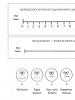
According to these documents, P. Beighton (1998) criteria should be used to diagnose HS:
— passive flexion of the metacarpal joint of the fifth finger by 90° in both directions (1–2 points, Fig. 1A);
- passive flexion of the first finger towards the forearm during flexion in the wrist joint (1-2 points, Fig. 1B);
- hyperextension of both elbow joints > 10 degrees (1-2 points, Fig. 1B);
- hyperextension of both knee joints > 10 degrees (1-2 points, Fig. 1D);
- when leaning forward with fixed knee joints, the planes of the patient's palms touch the floor (1 point).
The absence of hypermobility is determined with a score of 1 to 4, moderate HS - from 5 to 6 points, severe HS - from 7 to 9 points.
To make a diagnosis of SGS, one should use the Brighton criteria for SGS, which are divided into major and minor (Table 1).

GHS is diagnosed by the presence of two major criteria, either one major and two minor criteria, or four minor criteria. Two minor criteria are sufficient if there is a close relative with the disease. GHS is excluded in the presence of Marfan or Ehlers-Danlos syndromes. It is believed that the diagnosis of CGS in some patients today can be confirmed by laboratory studies of the level of tenascin X glycoprotein in the blood serum (muscle tendon antigen) and by analyzing the polymorphism of the tenascin X gene.
ICD diagnosis. In ICD-10, GHS has its own code and is assigned to class XIII - diseases of the musculoskeletal system and connective tissue, to block M30-M36 - systemic lesions of connective tissue and has code M35.7 - hypermobility syndrome. The ICD-10 also presents synonyms for HMS - family weakness of the ligamentous apparatus (Familial ligamentous laxity) and benign joint hypermobility (Benign joint hypermobility).
Diagnostic problems. Despite the existing diagnostic criteria, the proposed approach to diagnosis is not unambiguous. Since GHS is accompanied by lesions not only of the joints, and also taking into account the genetic aspects and the results of a number of morphological studies of fibrous structures of the connective tissue, some scientists have suggested that GHS and Ehlers-Danlos and Marfan syndromes be attributed to the same group of hereditary diseases located at opposite ends of the clinical spectrum. Today, the terms "joint hypermobility syndrome" and "hypermobility type of Ehlers-Danlos syndrome" are tried to be considered as synonyms, but a 2009 analysis of 3330 publications did not reveal reliable differential criteria for CGS, familial HS and hypermobility type of Ehlers-Danlos syndrome. The authors consider genetic characteristics to be the cause of SHS in children and propose a multidisciplinary approach to the management of children with HS and arthralgia with the aim of early initiation of preventive measures. In addition, to describe the combination of joint hypermobility and functional features, it is proposed to prefer to use the term "joint hypermobility syndrome", since the term "benign" in the description of "benign joint hypermobility syndrome" is misleading and inappropriate.
Difficulties in diagnosing SGS in children and adolescents also lie in the fact that the Brighton criteria were created for people from 16 to 85 years old and their use in pediatric practice may not be justified. Thus, epidemiological studies conducted among 6022 14-year-old children in the UK revealed 4 or more points on the Beighton scale in 27.5% of girls and 10.6% of boys. According to the authors, the obtained results cast doubt on the existing diagnostic criteria for HC in relation to young patients whose musculoskeletal system is in a state of growth and development, which requires clarification of the diagnostic algorithms for HC in children and adolescents.
Complications of GHS
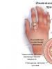
In the SGS clinic, in addition to the symptoms listed above, there are chronic pain syndrome with localization in typical places(Fig. 2). Such patients complain of constant pain in the shoulder and elbow joints, transient pain in the knee joints and in the lumbar spine, instability of the ankle and hip joints, clicks and subluxations of the joints, paresthesia in the femoral, calf muscles and distal phalanges of the hands, myofascial pain in the muscles of the shoulder belts.

We offer adapted from R. Keer, R. Gra-hame (2003) with additions to the classification of neuromuscular and musculoskeletal complications and clinical manifestations that are observed in children with joint hypermobility syndrome.
1. Acute or traumatic:
- sprain;
- damage:
Recurrent injuries of the ankle joint;
knee joint, meniscus rupture;
Acute and recurrent dislocations/subluxations:
Shoulder joint;
Patella;
Metacarpophalangeal joints;
Traumatic arthritis / synovitis;
Fractures.
2. Chronic non-traumatic:
- rheumatic affection of soft tissues:
tendovaginitis;
synovitis;
Juvenile arthritis/synovitis;
- pain in the knee joints;
- back pain (SHS is diagnosed in 55% of young people under the age of 30 with pain in the lower back);
- chronic widespread musculoskeletal pain syndromes;
- compression-radicular syndromes;
- flat feet and pain in the ankle joint;
- nonspecific arthralgia;
- scoliosis (SHS occurs in half of children with idiopathic scoliosis).
GHS doesn't just affect the joints. Thus, there is an opinion that one of the causes of postural tachycardia syndrome in persons aged 15-40 years and chronic daily headache syndrome is hypermobility of the spinal joints. Even psychiatric abnormalities, panic disorders, are more often detected in patients with SGS.
Results of own research
As part of the joint Ukrainian-Belarusian epidemiological study "Study of the health status and structural and functional state of bone tissue in children and adolescents who live in environmentally unfavorable regions", we examined 1259 students aged 10-17 from seven regions of Ukraine:
— Poltava region, town Mashevka — 231 children;
- Kharkiv region, Merefa - 240 children;
- Transcarpathian region, Kobyletskaya Polyana - 208 children;
- Donetsk region, Olenivka village - 88 children, Krasny Liman - 104 children, Mariupol - 98 children;
– Zaporozhye – 290 children,
and 594 schoolchildren from three regions of Belarus:
- Minsk - 205 children;
— Gomel region, urban-type settlement Lelchitsy — 194 children;
- Vitebsk region, Lepel - 195 children.
The examination program included a questionnaire based on a questionnaire developed by us, an objective examination by a pediatrician, anthropometry (42 indicators), assessment of sexual development, analysis of outpatient charts of child development and family history, ultrasound densitometry using the Sahara device (Hologic, USA), and, if necessary, the use of other instrumental methods (EchoCG, ECG, X-ray) and examination by narrow specialists.
Results:
1. The frequency of SGS registration among schoolchildren aged 10-17 living in Belarus was 11.2 ± 1.3%, in Ukraine - 16.8 ± 1.7%. The average score on the Bayton scale in children was 5.2-6.4, which corresponded to a pronounced degree of joint hypermobility.
2. Separate symptoms of connective tissue deficiency were diagnosed in relatives of almost half of children with SGS (40.5 ± 2.5% of children from Ukraine and 54.9 ± 2.9% of children from Belarus). Relatives of children with SGS were significantly more likely than in the group without GS to have varicose veins of the lower extremities, mitral valve prolapse and minor heart anomalies, myopia, and joint hypermobility. More than half of the mothers examined with SGS had pregnancy pathology, regardless of their place of residence (65.8 ± 2.3% in Ukraine and 69.0 ± 2.5% in Belarus). The data obtained testified to the indirect influence of heredity (manifestations of dysplasia in relatives) and the pathological course of the prenatal period of a child's development (preeclampsia) on the nature of connective tissue metabolism.
3. 40.5% of schoolchildren with SGS complained of headache, back pain, fatigue. In children with CGS, the dominant manifestations of connective tissue inferiority, in addition to joint hypermobility, were minor anomalies of the heart and pathology of vision, and impaired posture.
4. In girls with SGS, a delay in the start of sexual development was found in the age range of 10-14 years. From the age of 15, the pace of maturation leveled off. The sexual development of boys with SGS was characterized by a later start (from the age of 12) with a gradual leveling off of maturation rates.
5. SGS in 25% of schoolchildren was accompanied by a decrease in bone mineral density, while low physical activity as one of the causes of osteopenia was recorded in 3/4 of those examined with SGS, especially in Ukrainian schoolchildren (91-95% versus 69-78% in Belarus) .
Treatment
Given the polymorphism of CGS, the approach to managing such patients should be individualized. A decisive role in the treatment is given to lifestyle optimization and non-drug means, which includes the following activities:
1. Psychological support.
2. Selection of an adequate daily routine.
3. Diet therapy, which provides for a complete fortified diet with the appropriate amount of trace elements.
4. Physiotherapeutic methods, the basic principles of which in SGS were involuntarily formulated by D.V. Grigorovich in the story “The Gutta-perchie Boy”: “... After the first experiments, Becker (athlete-mentor. — Note. authors) made sure that he was not mistaken in the boy; Petya was as light as feathers and flexible at the joints; lacked, of course, the strength in the muscles to control these natural qualities; but it hasn't been a problem yet. Becker had no doubt that strength would be gained from exercise…” Indeed, physiotherapy and exercise in patients with CGS should be aimed at reducing pain, improving muscle strength, posture and proprioception, and correcting the movement of individual joints. Physical activity, which cannot be deprived of a child, must be dosed, meet the threshold of their tolerance for these patients. The complex of physical therapy includes isometric exercises, during which there is a significant muscle tension, but the range of motion in the joints remains minimal. Depending on the degree of manifestation of SGS, it is recommended to strengthen the muscles of the thigh and lower leg (knee joints), shoulder girdle, back, etc. It is easier for patients with SGS to achieve significant results in those sports that require flexibility, jumping ability, and small muscle mass from an athlete. Therefore, the most indicated for this category of patients are gymnastics, swimming, basketball and volleyball, choreography and dancing. If any complaints occur during sports, they should be temporarily stopped. Physiotherapy includes hydrokinesitherapy, massage and other techniques, depending on the degree of manifestation of hypermobility and the presence of other symptoms.
5. Carrying out vocational guidance for adolescents. Unfortunately, at present there are no normative documents on medical supervision and career guidance for adolescents with SGS. In our opinion, such patients should give up professions associated with prolonged standing, heavy lifting, vibration.
6. Depending on the clinical manifestations, children with SGS are recommended to be registered with a pediatrician who will work in close contact with narrow specialists (orthopedist, neurologist, dentist, etc.), draw up a treatment and rehabilitation plan for patients, monitor the effectiveness of measures.
7. Debatable methods of treatment of GHS. One of these is prolotherapy (Proliferative Injection Therapy) - one of the methods for treating the pathology of the ligamentous apparatus, which is used to stimulate reparative processes in it. The technique is used for pain syndrome associated with weakening of ligaments and tendons. The authors of the method claim that solutions injected into the places of attachment of ligaments and tendons to bone tissue promote the proliferation of damaged tissue. Various irritating solutions are used as proliferants, so the technique can also be called sclerosing therapy.
8. Drug therapy.
In addition to general principles, a number of therapeutic measures are used, which are determined by the characteristics of the course and the nature of the complications of CGS.
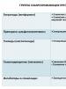
So, despite the low level of evidence (C or D), some authors recommend that patients with GHS take courses of basic drugs that directly or indirectly affect connective tissue metabolism:
- stimulants of collagen formation - vitamins C, B 1, B 2, B 6, folic acid, L-carnitine, trace elements (calcium, zinc, magnesium, manganese);
- correctors of violations of the synthesis and catabolism of glycosaminoglycans;
- stabilizers of mineral metabolism;
- local drug effect on the joints in the event of complications (plasters, ointments, etc.);
- surgical correction of deformities of the musculoskeletal system and chest.
9. Taking into account the data of a number of authors on the high (25-60%) incidence of osteopenia in children and adolescents with SGS, which may be associated with the peculiarities of remodeling processes or impaired mineral metabolism in this condition, one of the main directions in the treatment of SGS is the prevention of early osteopenic syndrome. .
Conclusion
Thus, in pediatric practice, the problem of SGS requires further study, since, on the one hand, the ability to distinguish between physiological and pathological HS appears only by the period of 18-30 years, on the other hand, the earlier SGS is diagnosed, the more effective will be measures to prevent the occurrence pathological symptoms caused by HS. The issue of prevention of complications and rehabilitation of children and adolescents with SHS, their medical and professional counseling remains insufficiently developed, which requires further research.
Bibliography
1. Isaev M.R. Clinical and epidemiological features of hypermobility syndrome in young people: Abstract of the thesis. dis... cand. honey. Sciences. - Orenburg, 2004. - 216 p.
2. Seckin U., Tur B.S., Yilmaz O. et al. The prevalence of joint hypermobility among high school students // Rheumatol. Int. - 2005. - Vol. 25, No. 4. - P. 260-263.
3. Korshunov N.I., Gauert V.R. Syndrome of hypermobility of the joints: clinical characteristics and features of rheumatoid arthritis and osteoarthritis that developed on its background // Ter. archive. - 1997. - T. 69, No. 12. - S. 23-27.
4. Keer R., Grahame G. Hypermobility Syndrome - recognition and management for physiotherapists. - Philadelphia, PA: Elsevier, 2003. - P. 177.
5. Grahame R., Bird H.A., Child A. The revised (Brighton 1998) criteria for the diagnosis of benign joint hypermobility syndrome (BJHS) // J. Rheumatol. - 2000. - 27(7). - 1777-9.
6. Kirk J.A., Ansell B.M., Bywaters E.G. The hypermobility syndrome. Musculoskeletal complaints associated with generalized joint hypermobility // Ann. Rheum. Dis. - 1967. - 26(5). — 419-25.
7. Beighton P., De Paepe A., Danks D. et al. International Nosology of Heritable Disorders of Connective Tissue, Berlin, 1986 // Am. J. Med. Gen. - 1988. - 29. - 581-94.
8. Loeys B.L., Dietz H.C., Braverman A.C. et al. The revised Ghent nosology for the Marfan syndrome // J. Med. Genet. - 2010. - Vol. 47(7). - P. 476-85.
9. Beighton P., De Paepe A., Steinmann B. et al. Ehlers-Danlos syndromes: Revised nosology, Villefranche, 1997 // Am. J. Med. Gen. - 1998. - 77(1). — 31-7.
10. Hereditary disorders of connective tissue. Russian recommendations. All-Russian Scientific Society of Cardiology — Section of Connective Tissue Dysplasia // Cardiovascular Therapy and Prevention. - 2009. - No. 8(6). — Annex 5.
11. Tofts L.J. The differential diagnosis of children with joint hypermobility: a review of the literature // J. Pediatric. Rheumatology. — 2009.
12. Clinch J., Deere K., Sayers A. et al. Epidemiology of generalized joint laxity (hypermobility) in fourteen-year-old children from the UK: a population-based evaluation // Arthritis Rheum. - 2011. - 63(9). — P. 2819-27.
13. Simmonds J.V., Keer R.J. Hypermobility and the hypermobility syndrome // Manual. Therapy. - 2007. - 12. - P. 298-309.
14. Stodolna-Tukendorf J., Stodolny J., Marczyński W. Spinal pain syndromes and constitutional hypermobility // Chir. Narzadow. Ruchu. Ortop. Paul - 2011. - 76(3). - P. 138-44.
15. Czaprowski D., Kotwicki T. et al. Joint hypermobility in children with idiopathic scoliosis: SOSORT award 2011 winner // Scoliosis. - 2011. - 7. - P. 6-22.
16. Mathias C.J., Low D.A., Iodice V., Owens A.P., Kirbis M., Grahame R. Postural tachycardia syndrome - current experience and concepts // Nat. Rev. Neurol. - 2011. - 6; 8(1). - P. 22-34.
17. Rubio-Agusti I., Kojovic M. et al. Cervical dystonia and joint hypermobility syndrome: a dangerous combination // Mov. Discord. - 2012. - 27(2). - P. 203-4.
18. Garcia-Campayo J., Asso E., Alda M. Joint hypermobility and anxiety: the state of the art // Curr. Psychiatry Rep. - 2011. - 13(1). - P. 18-25.
19. Garcia Campayo J., Asso E., Alda M., Andres E.M., Sobradiel N. Association between joint hypermobility syndrome and panic disorder: a case-control study // Psychosomatics. - 2010. - 51(1). - P. 55-61.
20. Hauser R.A., Phillips H.J. Treatment of joint hypermobility syndrome, including Ehlers-Danlos syndrome, with Hackett-Hemwall prolotherapy // Journal of prolotherpy. - 2011. - 3. - P. 2.
21. Podlianova O.I. Non-differentiation of vanadysplasia of the resulting tissue of hypermobility syndrome: breadth, specificity of diagnosing the disease: Abstract of the thesis. dis... cand. honey. Sciences: 14.00.09 // Simferopol, 2005. - 18 p.
22. Komrakova S.A. Bone mineral density in joint hypermobility syndrome in children: Abstract of the thesis. dis... cand. honey. Sciences: 14.00.09 // Ivanovo, 2006. - 18 p.
23. Benevolenskaya L.I. The problem of osteoporosis in modern medicine // Gynecologist. - 2005. - No. 12. - S. 10-15.
24. Juul-Kristensen B., Hansen H., Simonsen E.B., Alkjzhr T., Kristensen J.H., Jensen B.R., Remvig L. Knee function in 10-year-old children and adults with generalized joint hypermobility // Knee. - 2012. - P. 12.