Hypermobility of the joints mcb 10. Hypermobility of the joints - what is it, how to treat it? Treatment in the early stages
Connective tissue performs several functions in the human body at once. It is not responsible for the functioning of any organs, but at the same time forms their supporting frame and outer covers.
The organs of the human body are 90% composed of connective tissue. In some cases, a person may develop a special systemic connective tissue disease called dysplasia.
This term refers to a failure in the formation and development of connective tissue in humans. Dysplasia is a systemic disease and can involve groups of organs.
The disease can occur both at the stage of intrauterine development of the child, and develop after his birth.
The specificity of connective tissue dysplasia lies in the fact that it is not limited to only one specific manifestation, but is a group of diseases. Their feature is the non-inflammatory nature of the occurrence.
The syndrome is expressed as:
- damage to the structures and substance of the tissue;
- changes occurring in collagens, complex proteins, fibroblasts, elastic fibrils.
These defects become the main cause of violation of self-regulation in the body at any level, since the connective tissue is present in any part of it.
ICD designation
 For a long time there was no generally accepted name for this disease in medicine.
With the final confirmation of the systemic nature of the development of dysplasia, a general definition of the disease was officially approved - hypermobility syndrome.
This disease has an ICD-10 code - M35.7. Joint hypermobility according to the International Classifier is the main symptom of connective tissue diseases. This emphasizes the systemic nature of dysplasia.
In Russian medicine, a group of diseases is called connective tissue dysplasia. This term includes both syndromic and non-syndromic manifestations of the disease.
Reasons for development
The main provoking factor in the development of the disease are various gene mutations that the child's body undergoes during fetal development. Mutations affect various types of enzymes, protein-carbohydrate complexes.
There may be over 1000 different variants of genetic changes in proteins that provoke the development of the disease. The disease can be inherited.
Mutations are caused by the following factors:

With mutations, the following possible variants of disorders in protein chains can occur:
- their lengthening;
- truncation;
- development of selective mutations by substitution of amino acids.
Reference. It is assumed that one of the factors for the occurrence of connective tissue dysplasia in humans is insufficient intake of magnesium in the human body during embryonic development.
Symptoms
The manifestations of the disease are different. There are both light forms of it, and heavy ones that require a special approach. Symptoms and treatment of connective tissue dysplasia syndrome highly individual for each patient and in many ways unique.
The following manifestations of the disease are possible:

Symptoms depend on the type of disease. There are differentiated and undifferentiated forms. The signs of the first are:
- aortic aneurysm;
- fragility of bones;
- skin atrophy;
- finger deformity (arachnodactyly);
- scoliosis;
- funnel chest deformity;
- increased skin vulnerability (Ehlers-Danlos syndrome);
- Marfan's disease in the form of a violation of the shape of the skeleton, pathologies of the organs of vision and the cardiovascular system.
 The syndrome of undifferentiated connective tissue dysplasia is manifested by symptoms:
- increased skin elasticity;
- excessive joint mobility;
- skeletal anomalies;
- atypical thinness of the skin;
- various forms of malfunctions in the operation of myocardial valves, organs of vision.
Attention! People with undifferentiated dysplasia are not included in the number of patients, but belong to a group of patients prone to the manifestation of possible characteristic pathologies.
Diagnostics
The most accurate diagnosis can be established by the following methods:
- endoscope examination;
- skin biopsy;
- x-ray examination of the joints, lungs, spine;
- electrophysiological examination (ECG, electroencephalogram);
- blood test for biochemistry;
- Ultrasound of the kidneys and pelvic organs;
- medical genetic examination;
- daily urinalysis;
- measurement of body parts;
- joint mobility test.
The detection of problems in the functioning of several body systems indicates the likely development of connective tissue dysplasia in a patient.
Therapy Methods
Therapy for the disease should be complex and individual, depending on the symptoms and damage to the patient's specific body systems. Treatment of the disease includes:
- physiotherapy, performing special exercises;
- taking drugs to improve metabolism;
- compliance with the diet;
- surgical methods for deformation of the chest and musculoskeletal system.
Non-drug therapy includes:

Drug therapy includes taking the following drugs:
- metabolic stabilizers ("Alfacalcidol");
- collagen production stimulants (ascorbic acid, magnesium citrate);
- drugs that support the heart muscle ("Mildronate", "Lecithin");
- tissue repair stimulants ("Chondroxide");
- normalizing amino acid levels of drugs ("Glycine").
Patients need intensive nutrition. It is necessary to consume protein foods, fish, cheeses, seafood in large quantities. It is important to include meat-based broths, fruits and vegetables in the diet, and also take Omega-class dietary supplements.
Peculiarity! Surgical treatment is carried out only in two cases: when a person has a threat to life with severe vascular pathology and with obvious chest deformities.
Peculiarities of treatment in children
The syndrome of connective tissue dysplasia in children requires a special approach in its treatment. It is important to pay attention to the following methods:
- child's dietary intake(it should be dense and include various types of meat, legumes, fruits with vegetables, seafood);
- proper organization of life(refusal of serious sports loads in favor of physiotherapy and light gymnastic exercises);
- competent adaptation of the child to life in society(a lesson with a psychologist in order to prevent the formation of an inferiority complex);
- the use of special joint-strengthening splints and gypsum for young children;
- use of a course of metabolic stimulating drugs(the duration of the course is 60 days, after which a break is taken).
In case of serious pathologies against the background of the disease, the child needs surgical treatment in the form of a surgical operation. It is carried out with serious threats to the life of children with connective tissue dysplasia.
Important! Muscle dysplasia in children, as in adults, is not amenable to definitive treatment due to the genetic factor of its development. Therapy can only reduce the signs of its manifestation, slow down the symptoms or stop the development of the syndrome.
Contraindications
 If a person has this disease, the following is not recommended and prohibited:
- engage in hard and harmful work;
- perform exercises to stretch the spine or hang on the horizontal bar;
- expose yourself to stress and psychological overload;
- engage in contact sports, as well as weightlifting.
Conclusion
Connective tissue dysplasia syndrome is a group of diseases of genetic origin. They are characterized by a multiplicity of symptoms, which requires an integrated approach in diagnosis and treatment.
Taking into account the hereditary nature of the development of the disease, it is not amenable to final treatment, but the therapy used with it can significantly improve the quality of life of the patient and avoid the progression of pathologies up to the onset of old age.
Joint pain, arthritis accompany many diseases, follow them, or may precede a typical picture of an acute inflammatory process. Arthralgia with signs of local inflammation is characteristic of more than 200 diseases. It can be a leading symptom or one of the concomitant manifestations.
Arthritis (from the Latin artr - joint, itis - inflammation) - inflammatory lesions of the joints, differing in origin, localization, manifestations, but having common features of local inflammation and damage to the inner lining of the joint.
Among all rheumatological manifestations in childhood, reactive arthritis is the most common. In the older age group, it develops in young people under 40 years of age. In most manifestations, it is associated with acute intestinal infection caused by enterobacteria and acute urogenital chlamydial infection. May provoke the development of reactive arthritis and respiratory mycoplasma and chlamydial infections (Mycoplasma pneumoniae et Chlamydia pneumonia).
Reactive arthritis (ReA) is an acute inflammation of the joints of a non-purulent nature, symptoms develop no later than 1 month after an acute intestinal or genitourinary infection, associated with the histocompatibility antigen HLA-B27. It may be due to the development of mediated immunological inflammation after vaccination, with influenza, tuberculosis and other infections.

Thus, the true cause of the disease is not infectious inflammation provoked by the pathogen, but the damaging effect of immune complexes, which provokes a typical joint lesion with intra-articular fluid accumulation.
Classification in ICD-10
All of them belong to the class of infectious arthropathies: in the ICD-10 code M 00-M 03.
Code M 02 in ICD-10 - reactive arthropathies
Code M 02.0 in ICD-10 - arthropathy accompanying intestinal shunt
Code M 02.1 in ICD-10 - post-dysenteric arthropathy
Code M 02.2 in ICD-10 - post-immunization arthropathy
Code M 02.3 in ICD-10 - Reiter's disease
Code M 02.8 in ICD-10 - other reactive arthropathies
Code M 02.9 in ICD-10 - reactive arthropathy, unspecified
Classification of reactive arthritis (Table 1)
| Reactive arthritis | Working classification |
| By etiology | 1. Urogenital arthritis (most often caused by Chlamidia trachomatis). 2. Arthritis after intestinal infection. 3. Arthritis caused by another viral or bacterial infection. 4. Septic arthritis. Points 3 and 4 in practice are often combined by rheumatologists into the ReA group, although they are not such. |
| Flow | 1. Acute - up to 6 months. 2. Protracted - up to 12 months. 3. Chronic arthritis - more than 12 months. 4. Recurrent (the presence of a repeated attack after at least 6 months from the start of remission). |
| By degree of activity | 1. High. 2. Average. 3. Low. 4. Remission. |
| Development of functional insufficiency (FTS) | 1. Professional opportunity saved. 2. Lost professional opportunity. 3. Lost the ability to self-service. |
Most common location of joint lesions (Table 2)
| Causes of Arthritis | Typical joint damage |
| Dysentery | Symptoms of oligoarthritis of the lower extremities and sacroiliitis |
| Yersiniosis | Large joints of the legs, sacroiliac joints, calcaneus |
| Ulcerative colitis | Shoulder, hip, bilateral sacroiliitis, spondyloarthritis |
| Crohn's disease | Shoulder, elbow, sacroiliitis, spondyloarthritis |
| Gonococcal | Monoarthritis of the lower extremities |
| Reiter's disease | Knee, metatarsophalangeal, sacroiliitis spondyloarthritis |
| Tuberculosis | Hip, knee, spine |
| Brucellosis | Wrist, interphalangeal, ulnar, hip, knee, sacroiliac |
Symptoms
- Symptoms of general intoxication: fever from subfebrile numbers to high fever, general weakness is expressed, there is a decrease in appetite and weight.
- Symptoms of joint damage: asymmetrical reactive arthritis; characteristic is the defeat of both large and small joints of the legs - the ankle, knee and joints of the feet, especially the thumbs. The joints of the girdle of the upper extremities are less commonly affected: shoulder, sternoclavicular or temporomandibular, sacroiliac. At the same time, no more than six joints are affected.
- The development of inflammation in the areas of attachment of the joints and ligaments to the bones (enthesis). Most often, tendovaginitis of the toes or hands, the heel area develops.
- Mucosal lesions: symptoms of conjunctivitis with eye damage, urethritis and annular balanitis, cervicitis in women with damage to the genitourinary system, painful erosions on the oral mucosa.
- Signs of keratoderma: foci of hyperkeratosis of the plantar part of the feet or hands.
- Signs of damage to the nails (usually toes).
- Combined lesions of other organs:
- aortitis (inflammation of the wall of the aorta);
- myocarditis;
- mitral valve insufficiency;
- myositis - damage to skeletal muscles;
- polyneuritis - the appearance of symptoms of damage to the peripheral nervous system;
- swollen lymph nodes (for example, inguinal groups with urogenital pathology).
Additional methods for diagnosing arthritis
- Instrumental:
- radiography of the joint;
- spiral computed or magnetic resonance imaging;
- osteoscintigraphy;
- NMR spectroscopy;
- Ultrasound of the joint;
- arthroscopy.
- Laboratory:
- general clinical;
- biochemical research;
- immunological;
- immunoelectrophoresis;
- study of synovial fluid.
Information about what changes in the results of laboratory and instrumental examinations can be expected, we have systematized in Table 3.
| Diagnostic methods | Changes in ReA |
| Laboratory | |
| UAC | Decreased hemoglobin level, leukocytosis, thrombocytosis, increased ESR |
| Biochemical research | Increased CRP, hyperfibrinogenemia |
| Immunological study | An increase in the level of IgA, hypergammaglobulinemia, HLA-B27 in 60-80%. |
| Instrumental | |
| X-ray of the joint | Erosions, along with subchondral sclerosis, bone proliferation, osteosclerosis or osteoporosis with a protracted and chronic course, periostitis |
| Ultrasound of the joint | Cartilage thinning, thickening and deformation of joint surfaces, inflammatory intra-articular effusion, synovial hypertrophy, swelling of surrounding tissues |
| synovial fluid | Low density mucin clot, neutrophilic leukocytosis |
Differential diagnosis of reactive arthritis
Differential diagnosis of ReA is shown in Table 4.
| signs | Reiter's disease (urogenital reactive arthritis) | Rheumatoid arthritis | Systemic scleroderma | Psoriatic arthritis | Systemic lupus erythematosus |
| Floor | Predominantly male | 80% women | 80% women | Men and women with the same frequency | 90% women |
| Age | 18-30 years old | 10-55 years old | 30-50 years old | 20-45 years old | 30-40 years old |
| Start | Acute | Acute, subacute, chronic | gradual | gradual | Subacute |
| Antecedent factors | Symptoms of intestinal infection, sexually transmitted diseases, trauma | Viral infection, industrial and domestic chemical exposure, hypothermia, trauma, stress | Nervous strain | Viral infection, insolation | |
| Flow | Recurrent | rapid progression | slow progression | slow progression | slow progression |
| Symmetric joint damage | Not typical | Often | In 28% of patients | Seldom | Seldom |
| Frequent localization | knee joints | Interphalangeal proximal, wrist joints | Interphalangeal proximal joints, nail phalanges | Distal interphalangeal joints | Predominant damage to the periarticular tissues. Foci of necrosis of the femoral head, in the vertebral bodies, patella |
| morning stiffness | Not visible | Often | Not visible | Not visible | Not visible |
| Symptoms of damage to the skin and mucous membranes | Stomatitis, keratoderma of the hands and feet | Subcutaneous rheumatoid nodules. Atrophy of regional muscles | Puffiness with thickening and thickening of the skin of the face, spider veins | Psoriatic plaques, stomatitis, glossitis | Erythema of the face in the form of a "butterfly", erythema on the neck and dorsum of the hands, alopecia, brittle nails |
| Spinal injury | There is no pattern, but in the late stage, the lumbar region is more common | Rarely cervical | Not typical | No pattern, more often lumbar | No pattern |
| Symptoms of damage to other organs | Often urethritis, cystitis, conjunctivitis | Heart, kidneys, lungs | Lungs, heart, esophagus, kidneys | Skin, mucous membranes, rarely kidneys and heart | Heart (pericarditis), lungs (pleurisy), stomach, intestines, kidneys, nervous system |
Differential diagnosis of joint damage in reactive arthritis with other and articular pathology based on examination data is shown in Table 5.
| Reactive arthritis | The most common involvement of the knee and ankle joints, I toe; asymmetry of the lesion | Elevated ESR, mild leukocytosis, moderate thrombocytosis, anemia, CRP, pyuria, microhematuria, and proteinuria in urinalysis due to urethritis | Osteosclerosis, bone proliferation and marginal erosion, periostitis |
| Psoriatic arthritis | Damage to the interphalangeal joints, recurrent damage to the elbow, knee and ankle joints, pain is pronounced. May be malignant | Elevated ESR, slight leukocytosis, anemia, the content of fibrinogen and seromucoid is increased. Increased activity of acid phosphatase, proteinase, hyaluronidase. Presence of HLA antigen, complement | Subchondral osteoporosis and sclerosis, subchondral cysts, usuration of articular surfaces. Destruction of the epiphyses of the metatarsal bones. Sclerosis of intervertebral discs, change in their height |
| Rheumatoid arthritis | Stiffness after waking for more than 30 minutes. Swelling of the metacarpophalangeal, interphalangeal and radiocarpal joints. Flexion contracture of the fingers, ulnar deformity of the hand. Symptoms of atrophy of the muscles of the hands | ESR increased to 40-70 mm/h, the content of fibrinogen and seromucoid, ά2- and ɣ-globulins increased, the presence of CRP, specific rheumatoid factor (RF) | Destructive changes in the heads of II-III metacarpal and V metatarsal bones, bones of the wrist joint. Narrowing of interarticular fissures, cysts in the epiphyses of bones. Marginal bone growths, osteoporosis |
| rheumatoid arthritis | Symptoms of joint damage appear after suffering a sore throat, more often polyarthritis, volatility, symmetry of the lesion. Symptoms of simultaneous damage to the heart and joints. Subcutaneous nodules in the joints. erythema annulare |
Moderate leukocytosis, increased ESR, fibrinogen, seromucoids, ά2- and ɣ-globulins. The presence of SRP. An increase in the titer of ASL-O, IgM. | No changes, no joint disability |
| Systemic scleroderma | Deformation of small interphalangeal joints. Stiffness after awakening, flexion contractures of small, later large joints. Symmetry of the lesion | Anemia (B12-deficient, hemolytic or hypoplastic). ESR increase up to 25 mm/h. An increase in the content of fibrinogen, seromucoid. Increasing CRP | subchondral osteoporosis. The interarticular spaces are narrowed. Ankylosis |
There are three approaches to the treatment of reactive arthritis:
- drug treatment;
- functional treatment;
- treatment with folk remedies.
In the first case, the following means of therapy are distinguished:
- When a focus of infection is identified and the cause of arthritis is established, antibiotic treatment is carried out, taking into account sensitivity to the relevant microorganisms.
- NSAIDs are used to reduce signs of inflammation, pain intensity and hyperthermia.
- GCS systemically prescribed in case of severe systemic manifestations. More often, the treatment of corticosteroids is carried out in the form of intra-articular injections.
- The basic drug in the transition of arthritis to a chronic form is sulfasalazine for a long time (several months).
- Systemic enzyme therapy - treatment with Wobenzym.
Treatment with folk remedies involves both the use of decoctions and infusions of herbs with anti-inflammatory and antibacterial effects, as well as the local application of compresses from comfrey, horseradish, black radish.
Medications for medical treatment (Table 6)
| Preparations | Reiter's disease | Postimmunization arthropathy | Postdysenteric arthropathy | Pseudotuberculous arthritis |
| Doxycycline | 0.3 g 3 times a day | — | — | 0.3 g 3 times a day |
| Azithromycin | 1 g in 1 day, then 0.5 g | — | 1 g in 1 day, then 0.5 g | 1 g in 1 day, then 0.5 g |
| Ciprofloxacin | 1.5 g 2 r / d | — | 1.5 g 2 r / d | 1.5 g 2 r / d |
| Amikacin | — | — | 1 g/day | 1 g/day |
| diclofenac | 150 mg/day | 2-3 mg/kg/day | 150 mg/day | — |
| Meloxicam | 15 mg/day | 0.3-0.5 mg/kg 1 r/d | 15 mg/day | — |
| Levomycetin | — | — | — | 2 g/d |
| Celecoxib | 200 mg 1-2 r / day | — | — | — |
| Ibuprofen | 200 mg 2-3 times a day | 35-40 mg/kg in 2-4 doses | 200 mg 2-3 times a day | 200 mg 2-3 times a day |
| Prednisolone | 20-40 mg/day | — | 20-40 mg/day | — |
| Depo-Medrol | 0.1-40 mg / day | — | 0.1-40 mg / day | 0.1-40 mg / day |
| Diprospan | 2 mg / day | 1 ml IM once every 2 weeks | 2 mg/day | 1 ml IM once every 2 weeks |
| Sulfasalazine | Max. 2-3 g/day | 30-40 mg/kg | 0.5-1.5 g/day | 0.5-1.5 g/day |
| Phlogenzyme | 2 tab. 3 r/d | 2 tab. 3 r/d | 2 tab. 3 r/d | 2 tab. 3 r/d |
| Wobenzym | 5 tab. 3 r/d | 5 tab. 3 r/d | 5 tab. 3 r/d | 5 tab. 3 r/d |
Post-immunization reactive arthritis (after vaccination) develops more often in children, so it is necessary to adjust the dose of the drug per kilogram of the child's weight.
Similar symptoms can occur with arthritis of various etiologies. Only an experienced doctor can conduct a thorough diagnosis to determine the cause of arthritis and prescribe the correct treatment. It should be borne in mind that each drug has side effects and may be contraindicated in a particular case in a given patient. Even treatment with folk remedies should be carried out under the supervision of a physician; it is usually impossible to get rid of this disease completely, however, with adequate therapy, a long-term remission occurs. Forecasts for arthritis after intestinal infection are more favorable than for Reiter's syndrome, articular syndrome against the background of ulcerative colitis and Crohn's disease.
Sources:
- Attending doctor. E.S. Zholobova, E.G. Chistyakov. Reactive arthritis in children - diagnosis and treatment;
- VA Molochkov Moscow Regional Research Clinical Institute named after V.A. M.F. Vladimirsky, Moscow. Reiter's disease. Consilium Medicum. 2004; 03;
- V.M. Chepoy. Diagnosis and treatment of diseases of the joints. Moscow. "The medicine".
Joint hypermobility syndrome in children and adults: methods of treatment
Joint hypermobility is a condition that is characterized by an excess of the range of motion in the joint in comparison with physiological norms. The second name of the syndrome is connective tissue dysplasia. Hypermobility is considered a pathological condition, although it is not accompanied by inflammation or destructive-degenerative changes in tissues. But people with dysplasia are much more likely to develop joint disease.

Its early diagnosis (usually in childhood) will prevent premature destruction of the joints. Treatment of pathology does not require the use of drugs. Therapy is aimed at strengthening the joints, increasing the strength of the muscles and the ligament-tendon apparatus.
Development mechanism
The stable functioning of the human musculoskeletal system depends not only on the strength of the bones of the spinal column and limbs. The condition of the ligaments, tendons, synovial bags also matters. Connective tissue structures should be dense, but at the same time flexible and elastic. Under the influence of loads, such ligaments and tendons do not tear, but are slightly stretched. They protect the joint from damage, prevent injury.
Joint hypermobility is genetically determined. If parents during their life often tuck their ankles, their fingers unnaturally bend on their hands, then the child will inherit the same pathological structure of ligaments and tendons. Due to the peculiarities of metabolism, the synthesis of the most important bioactive substances, which are structural elements of connective tissues or take part in their synthesis, is disrupted. These include:
- collagen;
- proteoglycans;
- glycoproteins;
- some enzymes.
As a result of disruption of biosynthesis processes, the connective tissue loses its density and becomes excessively extensible. In most of the inhabitants of the planet, the state of the ligamentous-tendon apparatus is within the normal range, and only 10% of people are diagnosed with increased joint mobility.
Joint hypermobility is one of the characteristic features of Ehlers-Danlos syndrome, Marfan syndrome, osteogenesis imperfecta. If a person has a high extensibility of ligaments and tendons, differential studies are carried out to exclude pathologies.
Characteristic features of the syndrome in children
Hypermobility of the joints was previously considered not a pathology, but only a feature of the structure of the human musculoskeletal system. Flexible and plastic children parents sought to identify in different sections. It was believed that such a structure of the skeleton contributes to the rapid achievement of significant sports results. Currently, joint hypermobility in children is considered as a deviation from the physiological norm. A child with connective tissue dysplasia is contraindicated in certain sports:
- acrobatics and gymnastics;
- running and biathlon;
- football and hockey;
- long and high jumps;
- sambo, karate, judo.

During sports training, the joints of adults and children experience loads that exceed their strength limits. In people with a normal structure of joints, this can only cause injury - dislocations or sprains. After treatment, athletes quickly resume training. With hypermobility, events develop according to a different scenario. Any, even the most insignificant, injury can trigger destructive changes in cartilage, bone tissues, ligaments and tendons, and cause osteoarthritis.
Doctors advise parents of flexible and plastic children not to rush to take them to sports sections. Such a child needs a thorough examination. If he is diagnosed with hypermobility of the joints, then he will have to forget about athletics, strength sports, ballet and sports dancing.
Causes and provoking factors
Joint hypermobility is one of the symptoms of other diseases, but in most cases it is a genetic feature. A person does not even know about the need to correct such a condition, and sometimes treatment. In some cases, the syndrome is not inherited, but acquired during fetal development. Most often this happens in the first trimester of pregnancy, when the most important internal organs are laid in the embryo. The following adverse factors can provoke a breakdown in collagen production:
- women living in places with poor ecology;
- lack of proteins, fat- and water-soluble vitamins, microelements in the diet;
- infectious pathologies transferred during childbearing, especially of viral origin;
- frequent stress, depression.
Hypermobility syndrome is not provoked by internal or external factors (excess weight, excessive physical activity), which distinguishes it from most diseases. He himself becomes the cause of the development of pathologies.
Increased extensibility of ligaments and tendons leads to accelerated wear of articular structures, especially hyaline cartilage. Gradually, destructive-degenerative changes in the tissues occur, reducing the functional activity of the joints and causing the appearance of negative symptoms.
Clinical picture
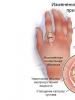
Many people, without even going to the doctor, realize that not everything is in order with their joints. This is indicated by frequent dislocations and subluxations, especially of the ankle. They try to minimize the chance of injury by avoiding heavy lifting and wearing low-heeled shoes. If a dislocation does occur, then almost always in a person with hypermobility, effusion accumulates in the joint cavity. In most cases, the synovial bag does not become inflamed, and the exudate is gradually removed from the joint. But painful sensations begin to arise when the weather changes, an acute experience of stress, in women during menstruation. The state of articular hypermobility is characterized by other pronounced symptoms:
- crepitus - specific clicks and crunch when walking or flexion-extension of the joint. For the state of hypermobility, it is not a sign of joint destruction, but occurs due to uneven sliding of the tendon relative to the bone protrusion;
- back pain, often in the lumbar region. May indicate the development of scoliosis and displacement of the vertebrae;
- development of symptomatic longitudinal, transverse or combined flat feet. It is more common in young women, accompanied by evening leg fatigue and the inability to wear high-heeled shoes;
- periarticular lesions. In patients older than 45, tendons and ligaments often become inflamed. The cause of the pathological process is excessive physical activity or long walking.

In patients older than 35 years, a symptom complex of joint hypermobility is often diagnosed. There are painful sensations, flat feet are complicated, injury to the ankles is becoming more frequent. This condition requires immediate medical attention, as it can lead to the development of arthrosis or arthritis.
Symptoms of general intoxication of the body appear with the development of synovitis, or inflammation of the synovial sac after injury. The patient's body temperature rises, digestion is upset, severe headaches occur. There is a possibility of infection of the joint with pathogenic bacteria.
Diagnosis and treatment
An experienced diagnostician is able to detect pathology by extra-articular signs of hypermobility. High extensibility of the ligaments is indicated by skin without fatty layers, long thin fingers, height above average, lean physique, broken dentition. These features of the organism are based on the specific structure of connective tissue structures. Questioning the patient helps to make a diagnosis: he complains of frequent injury, a predisposition to bruising after a slight external impact. To differentiate the syndrome of hypermobility of the joints from arthritis, osteoarthritis, coxarthrosis, gonarthrosis, a number of instrumental studies are carried out:
Joint treatment More >>
- radiography;
- magnetic resonance or computed tomography.
Their results also make it possible to determine the degree of damage to the tendon-ligamentous apparatus, the number of complications that have developed.

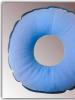
Treatment is required only with the development of articular pathologies provoked by hypermobility of the joints. In all other cases, the patient is recommended to strengthen the muscular corset and ligament-tendon apparatus: engage in physiotherapy exercises, swimming or just walk in the fresh air. Wearing orthopedic devices helps to relieve stress on problem joints:
More
- elastic bandages;
- posture correctors;
- interdigital pads.
People with such a structure of ligaments and tendons should avoid wearing high-heeled shoes, be careful when moving over uneven terrain. Under the ban - active sports training, in which the joints are often injured.
Dislocation of the hip endoprosthesis: symptoms and treatment after endoprosthesis replacement
Sometimes, due to the characteristics of the body, the patient has certain complications after hip arthroplasty. The most common violation of the full functioning of the limb is dislocation of the endoprosthesis head.
Since an artificial joint cannot completely replace natural tissues, for this reason its functionality is reduced. In this regard, any careless movement of the hip joint, very early rehabilitation or any difficult exercise can cause dislocation of the endoprosthesis. Including a normal fall can lead to this.
Symptoms of dislocation of the hip endoprosthesis
Dislocation of the hip endoprosthesis is a violation of the contact of the femoral head with the acetabular component, in which case emergency reduction is required.
Due to certain characteristics of the body, the following are primarily predisposed to dislocation of the artificial hip joint:
- Patients diagnosed with hip fracture and dysplasia;
- Patients who have undergone previous surgery;
- Patients with hypermobility of the hip joint.
The symptoms of dislocation of the endoprosthesis are similar to the symptoms of violation of healthy joints. In particular, the patient feels a sharp pain when walking and at rest, weakness in the lower extremities, and the support ability of the artificial hip joint decreases.
A swelling forms around the broken joint, while the lower limb is visually shortened. If you do not consult a doctor in time and start surgical treatment, the patient's body temperature may rise sharply due to the activity of the inflammatory process.
Why is a dislocation of the hip joint formed?
Risk factors for endoprosthesis dislocation can be divided into three large groups: patient-related, implant design-related, and surgeon-controlled. In the period after the operation, if the rules are not followed and careless movements, the patient may experience a complication in the form of a violation of the prosthesis.
Dislocation of an artificial hip joint can be caused by all sorts of reasons. This may be a human factor, when the patient himself is to blame for what happened. Also, a violation can occur due to the poor quality of the endoprosthesis. The mistake of a surgeon with a lack of personal experience is not excluded.
The main reasons may be:
- Poor contact of articular surfaces;
- Poor-quality installation of the endoprosthesis;
- Excessive load on the artificial joint after the operation;
- Excessive body weight of the patient;
- The occurrence of shearing or torque;
- Infection in the joint cavity;
- Abrasion of the joints.
Including dislocation can be formed with a fracture of the neck, osteoporosis, aseptic necrosis of periprosthetic bone tissues. Violation of the anatomy of bones and muscle function.
A fairly high risk of dislocation in the elderly. According to statistics, it is people over 60 who most often come with such complaints after joint replacement surgery.
Since women have a large initial range of motion in the hip joint and less muscle mass, they are primarily predisposed to malfunction of the prosthesis. Including tall people with growth above average fall into the risk group.
Risk factors associated with implants include the type of endoprosthesis, which can be unipolar, bipolar, dual mobility, and so on. The quality of the endoprosthesis depends on the type of stem and features of its design. The geometric parameters of the liner, head size, type of friction pair are also taken into account.
In particular, the liner in the form of an anti-luxation lip, which increases the degree of overlapping of the head with polyethylene, contributes to the increase in the "jump distance" of the head of the hip joint endoprosthesis. Also, the amplitude of movement depends on the size of the head - the higher it is, the greater the "jump distance".
The neck of rectangular cross-section allows for a greater range of motion in the joints.
Treatment of impaired mobility of the hip joint
In the event that the patient complains of the above symptoms, the doctor prescribes an X-ray examination. If a dislocation of the implant head is detected, an emergency closed reduction is performed under anesthesia or spinal anesthesia.
The nature of the operation depends on the reason for the dislocation, it can vary from open reduction and lengthening of the neck to replacement of the type of endoprosthesis.
After treatment, the patient is shown bed rest for 7-10 days. Next, you need to visit the physiotherapy room to strengthen the abductors and muscles of the anterior group. The patient is re-learned to walk under the supervision of a physiotherapist.
As a means of immobilization, a derotational boot, a back splint on the knee joint or a gypsum bandage are made.
How to prevent dislocation of the joint after arthroplasty
In the first days after the operation, the patient can sit down and get up only in the presence of a doctor or an instructor in therapeutic exercises. In any position, the operated leg should be no closer than the line of the imaginary continuation of the spine.
Do not make rotational movements, especially to the outside. For this reason, all turns must be made towards the operated limb. You should not heavily load and strain your leg, step on it with all your weight.
After a few weeks, the load on the joints can be gradually increased, but at this point the patient should use a cane. To prevent unwanted movements, the bed must have the required height, it is also important to properly equip the apartment.
After six weeks, the patient can gradually return to their normal routine. To prevent a violation of the functionality of an artificial implant, after arthroplasty, you should follow the basic rules.
- First of all, it is important to remember the right angle rule. It is impossible to bend the legs in the hip joints by more than 90 degrees, all movements must adhere to the amplitude of the right angle. It is also not recommended to cross your legs and squat down. In order not to forget about this rule, it is worth using special soft pillows that are placed between the legs.
- After sleep, you should only sit on a chair or chair with a straight back so that the flexion in the hip joints during sitting is less than 90 degrees. When getting up from a chair, the back should be straight, not leaning forward. You need to sit down with your legs slightly apart.
- While lying or sitting, it is recommended to move the operated lower limb slightly to the side. To control the correct position, you should follow the rule of thumb. In particular, the thumb is placed on the outer surface of the thigh, and in this position the knee should be further than the thumb.
- While in bed, you do not need to pull the blanket over yourself, lying at your feet. To do this, you can use some kind of additional device or simply ask someone to straighten the blanket. Similarly, you can not put on shoes without a spoon.
These basic rules must be followed after the operation at an early stage of rehabilitation. If the rehabilitation goes without consequences, the restrictions in movements will gradually disappear.
It is important to understand that the prosthesis is not a new healthy joint, but a mechanism that allows you to live and move without pain. After some time, it wears out, the average life of simple models is about 20 years. The speed of wear, in turn, depends on the patient himself.
It is necessary to avoid lifting heavy objects, standing for a long time, jumping. You should watch your own weight. Handrails must be used when going up and down stairs. Shoes should be low-heeled with non-slip soles.
In order to timely detect any abnormalities in the work of artificial joints, it is important to take regular control pictures and visit a doctor for a consultation.
How to prepare an apartment after surgery
After the patient is discharged and goes home, he usually encounters some difficulties during the performance of normal household tasks that were previously solved without problems. These difficulties can be avoided if the apartment is prepared in advance while the patient is being treated.
If a carpet is laid on the floor in an apartment, it is better to remove it for a while. It is important that the floor is level, as patients may cling to the edge of the carpet with their feet or a support with which they move after surgery.
On the walls in different places you need to place special strong handrails - they will come in handy in the bathroom, toilet, kitchen, next to the bed.
If possible, it is desirable to install a special medical bed, which allows you to change the height, provides additional safety and convenience when the patient gets on and off. The patient will be able to accommodate quite comfortably.
In the bathroom, when washing, you need to use a special wooden board for sitting; for a shower cabin, a chair with non-slip legs is suitable. Handrails should be installed on the walls of the bathroom so that the patient can freely and without problems enter, exit, sit down and get up safely.
After the operation, the patient in the toilet, the standard height of the toilet bowl will be small, so a special device will be required. To achieve the desired height and comfort, nozzles are usually used. Additionally, handrails should be installed in the toilet so that it is convenient to sit down and get up.
The video in this article will show how the endoprosthesis is installed, and how the patient's life changes after such an endoprosthesis.
This information is intended for healthcare and pharmaceutical professionals. Patients should not use this information as medical advice or recommendations.
Pathology of the spine with hypermobility of the joints
PhD A.G. Belenky, corresponding member RAMS, Professor E.L. Nasonov
RMAPO
Generalized joint hypermobility (GMS) is a condition that occurs in 10-15% of the population and is characterized by excessive (compared to the average in this age and sex group) range of motion in the joints. HMS is often found in members of the same family and tends to be inherited through the female line. Actually, HMS is not a pathological condition, but it is known as a reliable risk factor for both non-specific complaints from the musculoskeletal system, and for morphological signs of “weakness” of connective tissue structures of other body systems (heart valve prolapse, nephroptosis, varicose veins, prolapse of the uterus and etc.). The morphological substrate underlying the pathological signs is the greater than normal extensibility of collagen, which is present everywhere in the body. In a pronounced form, the signs of "failure" of connective tissue structures arising and accumulating during life form a clinical picture. hypermobility syndrome (GS) (code M37.5 according to ICD-10), which has its own diagnostic criteria.
The list of structures involved in the pathological process in both symptomatic HMS and HS naturally includes the spine. Like other forms of pathology of the musculoskeletal system in HMS and HS, spinal lesions are represented by a group of conditions, diseases and syndromes, united by a primary non-inflammatory genesis and distinct family aggregation. This list includes: nonspecific dorsalgia, scoliosis, Schuerman-Mau disease, spondylolisthesis and early osteochondrosis. None of the listed conditions is pathognomonic for HMS, which does not allow them to be used as the main criteria for the syndrome, however, many studies have shown that these types of spinal pathology are significantly associated with HMS.
Currently, when there are already developed criteria for the syndrome, HS largely remains a diagnosis of exclusion, that is, the condition is the absence of signs of other rheumatic diseases. However, despite the obligatory nature of such a "negative" component, the list of small "positive" criteria for HS includes spinal damage in the form of "dorsalgia for 3 months or more." As a separate minor criterion, spondylolisthesis is present. The inclusion of spinal involvement in the latest (Brighton) criteria for HS (1998) was a step forward in the process of refining the clinical manifestations of HS, which began with the pioneering work of Kirk et al. (1967) who determined the importance of HMS as a reliable cause of rheumatic pathology. The inclusion of spinal lesions in the additional criteria for GS was the result of clinical observations that showed a close relationship between HMS and spinal pathology, including in patients meeting the GS criteria. A feature of the listed lesions of the spine in HS is the possibility of their detection in isolation, in the form of separate nosological forms. But most of the authors who have studied non-inflammatory and non-traumatic spinal pathology indicate, on the one hand, a clear family accumulation of these conditions; on the other hand, the undoubted connection of the indicated pathology of the spine with other signs of systemic connective tissue dysplasia. The latter include foot deformities (longitudinal and transverse flat feet, "hollow" foot) and minor skeletal anomalies (deformities of the chest, toes, feet), known as phenotypic signs of connective tissue dysplasia. The list of the latter also includes HMS. In other words, non-inflammatory diseases of the spine, which debut in childhood and adolescence and have a distinct hereditary component, can be considered as a particular manifestation of a general pathological process. Such a view on the problem of early non-inflammatory pathology of the spine allows a doctor (primarily an orthopedist and rheumatologist) to put into practice the well-known principle of medicine - "to treat not the disease, but the patient."
In 20-50 years. of the last century, the problem of “dysraphic status” nosologically close to HS was actively discussed in the medical scientific literature. The latter was understood as a combination of various congenital developmental anomalies, mainly of the skeleton and nervous system. However, despite the undoubted relevance of the problem, the efforts made have not led to the creation of a unified system of views on the pathology under study. The reason was the disagreement of the authors on the question of what should be considered signs of dysplasia. Later, in the 50-60s. In the twentieth century, when developing the classification of scoliosis, its form was distinguished, defined as "dysplastic scoliosis", i.e. scoliosis, combined with other signs of skeletal dysplasia - flat feet, HMS, large and small phenotypic anomalies of the skeleton. However, in the future, due to the lack of differences in the clinical manifestations of scoliosis itself, in the prognosis and approaches to treatment, the separation of dysplastic and idiopathic scoliosis was considered inappropriate.
These historical facts indicate a periodic rise in interest in the problem of the relationship between early pathology of the spine and other signs of connective tissue dysplasia. However, due to the absence of pathognomonic symptoms, the variability of clinical manifestations and, most importantly, the absence of biochemical and genetic markers of these conditions, the solution to this problem was seen only in the future.
The current stage of the state of the problem of connective tissue dysplasia looks promising. On the one hand, the search for biochemical markers in certain stable combinations of clinical signs does not stop (there are successes here: individual subtypes of Ehlers–Danlos syndrome have been genetically characterized; genes responsible for the development of Marfan syndrome and osteogenesis imperfecta have been found). On the other hand, clinical observations made it possible to dwell on HMS as a universal sign of connective tissue dysplasia. Really, HMS is an easily defined clinical sign that reflects the state of not only the musculoskeletal system, but the entire connective tissue matrix. This approach is implemented in the international recognition of the term "hypermobility syndrome", which currently most fully characterizes the state of undifferentiated connective tissue dysplasia. On the one hand, the name indicates generalized joint hypermobility as an important clinical sign; on the other hand, the absence of the word “joint” in the definition reflects the complexity of the problem, which is not limited to the musculoskeletal system.
The most common manifestation of spinal injury in HMS is dorsalgia . Of course, this is a symptom, but not a diagnosis. In the population (especially in older age groups) this is the most common complaint from the musculoskeletal system. According to our studies (800 adults in the Moscow population aged 16 to 50 years), dorsalgia occurred with a frequency of 12% (among men 16–20 years old) to 35% (among women 41–50 years old). Among persons with HMS, the prevalence of dorsalgia is much higher - from 35% among men aged 16–20 to 65% among women aged 41–50. Qualitative differences in dorsalgia among hypermobile persons consisted in a significant predominance of thoracalgia in comparison with non-hypermobile persons, in whom lumbalgia predominated. In most cases, X-ray examination did not reveal any structural causes of dorsalgia. Clinical manifestations of dorsalgia in HMS are nonspecific - pain appears or increases with prolonged static loads (standing, sometimes sitting), decreases or disappears in the supine position, as well as with adequate treatment, including the use of centrally acting muscle relaxants, analgesics or non-steroidal anti-inflammatory drugs (NSAIDs), massage and gymnastics that strengthens the paravertebral muscles. It should be noted that true inflammatory diseases of the spine (occurring in the population with a frequency of 0.1–0.2%) can also be the cause of dorsalgia in people with HMS. In this case, another is observed - an inflammatory rhythm of pain with a maximum at night and in the morning and a more pronounced effect of NSAIDs. The possibilities of using NSAIDs in the differential diagnosis of the causes of dorsalgia and arthralgia are known. In terms of correction of dorsalgia in HMS, central muscle relaxants play a very important role. Their use allows, on the one hand, to achieve a more pronounced therapeutic effect, and on the other hand, to reduce the daily dose of NSAIDs and, accordingly, reduce the risk of developing NSAID-associated adverse events. Among muscle relaxants of central action, it has proven itself well. tolperisone (Mydocalm) , which has been successfully used for many years in many diseases accompanied by an increase in muscle tone. The daily dosage of Mydocalm in most cases is 450 mg (divided into 3 doses), the duration of taking Mydocalm depends on the patient's condition. The effect of including Mydocalm in the drug complex is not only to reduce the pain syndrome, but also to increase the range of motion. The latter circumstance affects another important aspect in the prognosis of the course and correction of dorsalgia, namely, the possibility for the patient to perform a physical rehabilitation program. It is well known that the more carefully a patient follows the recommendations for physical rehabilitation, the better his functional prognosis. Accordingly, a decrease in reflex muscle spasm allows, when performing physical exercises, to achieve a greater range of motion in the spine.
The second most common spinal lesion in HMS is scoliosis . In the population, scoliosis occurs with a frequency of 5–7%, does not differ by sex, and usually develops in childhood. The degree of scoliosis does not tend to increase after adolescence. Often there is asymptomatic scoliosis (up to 30 years), but the presence of thoracalgia is more typical. According to our data, the incidence of scoliosis in HMS is 30–35%. The pain syndrome in scoliosis is nonspecific and corresponds to the above description of dorsalgia in HMS, but is more pronounced and persistent. Orthopedic care should be provided as early as possible; It is known that after adolescence (and in some cases even with timely intensive treatment) there is no cure. The main role in the correction of scoliosis belongs to physical methods of influence. However, it is advisable to supplement rehabilitation programs with the use of muscle relaxants, and, if necessary, also with analgesics or NSAIDs. This can significantly improve both the quality of life and the patient's ability to participate in a rehabilitation program.
Spinal osteochondropathy described by H.W. Scheuermann in 1920, as aseptic necrosis of the apophyses of the vertebral bodies, in the ICD-10 is attributed to juvenile osteochondrosis. The prevalence of Schuerman-Mau disease (according to radiological signs) in the population is 2-5%. In the study of Maslova E.S. the presence of this pathology was shown in 11% of patients with HS (almost always associated with clinical kyphoscoliosis) and in 2% of non-hypermobile individuals in the control group. Clinical manifestations schoerman-mau disease do not differ in specificity and correspond to the above-described picture of dorsalgia in HMS, differing only in resistance, a tendency to lifelong preservation of spinal deformity and the development of radiological signs of secondary osteochondrosis at a young age. The principles of therapy for Schuerman-Mau disease are to start as early as possible, use methods that correct posture, optimize lifestyle (sleep on a hard bed, lifelong therapeutic exercises, including sports that strengthen the dorsal muscles - tennis, swimming), back muscle massage. As with symptomatic scoliosis, the course use of muscle relaxants is periodically indicated, and if necessary, NSAIDs are used as symptomatic therapy.
Spondylolisthesis (persistent displacement of the vertebral bodies in the horizontal plane) is most logically united by the common pathogenesis with HMS. One of the causes of spondylolisthesis is the increased extensibility of the powerful ligamentous apparatus of the spine. Another factor that stabilizes the position of the vertebrae is the condition of the facet joints. Apparently, the relative rarity of detection - 0.5–1% (compared with other types of spinal pathology) - of spondylolisthesis in HMS is associated with the latter. Despite the rarity, this spinal lesion in HMS is the most specific, which is reflected in the inclusion of spondylolisthesis as a separate feature in the diagnosis of HS. Spondylolisthesis in HS may be accompanied by signs of persistent mechanical radiculopathy and require prompt stabilization of the affected vertebral segment.
Thus, spinal lesions in HMS can manifest themselves in various types of pathology, differing in the severity of clinical symptoms, prognosis, and, to a lesser extent, treatment approaches. The general principles of therapy for a patient with HS are as follows:
1. The complexity of the approach, i.e. a look at all the patient's health problems (not only with the musculoskeletal system) through the prism of a possible generalized "failure" of the connective tissue. Often, this approach allows you to combine pathological manifestations from various body systems with one cause and one diagnosis.
2. Particular attention is paid to non-drug methods of treatment and rehabilitation.
3. Explanation to the patient of the need for long-term, sometimes lifelong, compliance with recommendations aimed at correcting and preventing further progression of spinal deformity, increasing and maintaining the strength of the paravertebral muscles.
4. Symptomatic treatment (analgesics or NSAIDs) should be used with caution (danger of side effects).
5. For pathogenetically-oriented drug therapy of pain in HS, central muscle relaxants (Mydocalm) are used.
Literature:
1. Beigton P., Graham R., Bird H. Hypermobility of joints. //2-nd edition. London, Berlin, Heidelberg et al. - Springer-Verlag. – 1989 – 189 p.
2. Belenky A.G. Generalized joint hypermobility and other connective tissue syndromes (review) Scientific and practical rheumatology. 2001. No. 4., pp. 40–48
3. Maslova E.S. Age features of clinical manifestations of joint hypermobility syndrome. // Diss... cand. honey. Sciences. - Moscow. - 2002. - p.152
4. Kirk JH, Ansell BM, Bywaters EG. The hypermobility syndrome // Ann Rheum Dis - 1967 - v.26.- p. 425–427.
5. Nikitina T.I. Clinical and genetic analysis of dysplastic scoliosis.// Diss. cand. honey. Sciences. Moscow. 1991.– p.1–234.
6. Davidenkov S.N. On the theory of the dysraphic genotype. Soviet neuropathology, psychiatry and psychohygiene. 1925, Vol.6.
7. Kaz'min A.I., Kon I.I., Belen'kiy V.V. Scoliosis. // M. Medicine. - 1981. - p. 272.
8. ICD 10. International Statistical Classification of Diseases and Related Health Problems. Tenth revision.// WHO. Geneva - 1995. - Volume 1., Part 3. - P. 665.
9. Belenky A.G. Non-steroidal anti-inflammatory drugs as a tool for differential diagnosis in articular syndrome. Russian Medical Journal, 2003, Vol. 11, No. 15 (187), p. 886–888
10. Lila A.M. Osteochondropathy.// Clinical rheumatology./Ed. Mazurova V.I. - St. Petersburg: Folio, 2001. - P. 372-381.
The most complete answers to questions on the topic: "hip dysplasia, ICD code 10".
Congenital diseases of the musculoskeletal system are becoming more common, and among them is hip dysplasia. Due to the wrong lifestyle of the mother during pregnancy, bad habits, as well as against the background of taking certain medications, children are born with underdeveloped limbs, cartilage and musculoskeletal systems. One of the most unfavorable outcomes is congenital dislocation of the hip.
In the international classification of diseases, this disease is allocated a separate class and group: M24.8 - a specified lesion of the joints that has not been displayed and is not classified in other groups.
What is the most common cause of this condition?
Pathology of joint development in a newborn
In newborns during fetal development, underdevelopment of the articular surfaces that form the hip joint is observed, which can lead to the disease congenital hip dysplasia.
The acetabulum is represented mainly by cartilage tissue. During the period of intrauterine development and neonatality, its cavity increases due to the cartilaginous lip (limbus). The cavity itself consists of three bones, the final ossification of which ends by the age of 18. The head of the femur, its neck and greater trochanter consist mainly of cartilage (these formations are not visualized on the radiograph). The head of the femur in newborns is always larger than the articular surface of the pelvis. All of the above factors cause insufficient joint strength.
The discrepancy between the articular surfaces (discongruence) creates conditions for the development of hip predislocation - a condition in which spontaneous dislocation of the femoral head occurs, its reverse reduction (this does not require much effort). In some children, spontaneous repositioning of the head of the bone occurs, after which the joint develops normally. In others, due to a long-term pre-dislocation, the head of the bone is displaced relative to the articular cavity, after which subluxation of the femur develops (the femoral head is displaced, but it does not go beyond the boundaries of the limbus).
If such a condition exists for quite a long time, then over time, due to the traction of the muscles gaining tone, the limb is pulled up and to the side (which one depends on the displacement of the head), due to which a dislocation of the femur develops. Gradually ossified, the head is fixed in the region of the anteroinferior pubic bone, receiving a new fulcrum there (in severe cases, the displacement of the head can occur in the region of the posterior surface of the iliac wing). In this case, there is a decrease in the relative length of the limb.
Typically, children develop a type 2a hip joint. There are several types of joints that correspond to the stages of development of a dislocation:
- 1 type - a healthy joint;
- type 2 - violation and slowing down of ossification;
- type 3 - hip subluxation;
- Type 4 - complete dislocation.
Type 2a (develops in babies up to 3 months old) is characterized by the presence of a rounded and short bone roof of the acetabulum, alpha and beta angles within 55 degrees (these angles characterize the position of the head along certain landmarks or axes), as well as a centered bone head without displacement. When the process is started, a step-by-step transition to dislocation is observed.
At the heart of the development of this pathology, the decisive role belongs to the incorrect position of the fetus during pregnancy. As statistics show, most often hip dysplasia develops with breech presentation with arms and legs crossed in front. This is usually observed in girls, especially with the left-sided position of the fetus in the womb.
Due to the adoption of such a position of the body, the heads of the femoral bones are displaced relative to the joint cavity, which is why hip dysplasia develops. With a long stay in this position, the cartilage of the bones cannot fully develop (since this requires their full compliance).
Diagnosis begins immediately after the birth of the child. Already in the maternity hospital, in order to establish dysplasia, it is necessary to determine the presence of a “click symptom” - this symptom manifests itself only with a dislocated joint (which can occur as a result of improper delivery and the help of a midwife to the baby). The symptom is determined due to the fact that the baby’s joint capsule is overstretched, due to which the femoral head “walks”. A click is detected after it slips off the edge of the acetabulum. This symptom, which indicates hip dysplasia, is checked very carefully so as not to damage the baby's delicate tissues and blood vessels.
To identify the symptom, the baby is laid on the table, his legs are bent at the hip and knee joints. The thumb is located on the inner side of the baby's thigh, and the remaining four are on the outer surface of the joint (in this case, the middle finger of the hand must be on the greater trochanter of the femur). The baby's hip is retracted to the side at a slight angle (usually up to 40 degrees), after which the child's hip is traction along its axis. In parallel, pressure is applied to the greater spit. With the correct procedure, you can feel a loud click (it is not perceived by hearing, but is felt due to tactile sensations).
Standard diagnostics for patients with pathology of joint development
As you grow older, this symptom loses its value, and others come to replace it. First of all, it becomes possible to determine the increased tone of the muscles of the limb.
This symptom should be identified together with neurologists to differentiate dislocation from neurological pathology. The symptom is defined as follows. The baby is carefully laid on the table. Putting your hands on the inner surface of the child's thighs, they abduct his hips to the sides. Normally, the child's hips should lie on the surface of the table (for a newborn); if the child is older, then the angle between the surface of the table and the leg should be at least 60 degrees. This restriction develops due to high muscle tone, and also, at a later age, due to the fact that the femoral head rests against the ilium.
With unilateral dislocation, an important place is occupied by a symptom of a violation of the symmetry of the longitudinal axes. With the legs bent at the knee joints, when they are moved to the sides, a visual decrease in the longitudinal axis of the limb is observed (due to the fact that the head is not in the articular cavity, but is shifted away from it). In parallel, on the side of the dislocation, there is a retraction of soft tissues in the area of the Scarpa triangle. Together with them, asymmetry of skin folds is observed.
There are several signs of hip dysplasia. At the age of one year, one of the main symptoms of the development of dislocation is diving lameness (with unilateral dislocation) or duck gait (with bilateral lesion).
In addition to determining the gait, the Trandeleburg symptom is also detected: when standing on a diseased leg with a healthy one raised and bent at the knee, the body shifts towards the healthy leg with a parallel retraction of tissues in the region of the diseased joint. There is a lowering of the gluteal fold.
Wrong gait in the pathology of the development of the hip joints
Additional symptoms are displacement of the greater trochanter above the Roser-Nelaton line and increased curvature of the lumbar spine.
X-ray diagnostics for children under one year old is not carried out, since the cartilaginous structures do not delay x-rays, and they pass by the bone. For a more reliable diagnosis, ultrasound (ultrasound) is used.
X-ray becomes possible to use from the age of one (by this age the femoral head is already ossified), and it is already possible to determine the displacement of the femur.
To determine the degree of displacement of the head, special schemes are used. The most widespread among them was the Hildenreiner scheme. It evaluates the spatial arrangement of the structures of the hip joint. In addition, additional schemes can be used, but the essence of their use is one thing: they all allow you to determine the position of the displaced femoral head and identify the degree of displacement and the possibility of active movements in this joint.
Arthrography and tomography of the joint provide the most complete and reliable information regarding the joint.
There are currently no specific treatments for dysplasia. If the defect was discovered during the neonatal period, most often the displaced femoral head is set back into place and the joint begins to develop normally on its own. If the condition has been started, resort to the correction of the displaced limb. To do this, use the so-called wide swaddling for dysplasia or setting up a spacer.
Swaddling for dysplasia is applicable in young children (most often used before the age of 4 months). Due to the tight swaddling of the legs, they are given a forced position of abduction, in which the articular head returns to its place.In children who have reached the age of six months, tight swaddling, Freik's pillow or the use of Pavlik's stirrups will no longer have the desired effect. In order to correct the joint, they begin to use the gypsum of the limb according to Ter-Egizarov. In parallel, spacers are placed in the area of \u200b\u200bthe knee joints. As the joints straighten and muscle tone decreases, abduction of the limb increases, which allows for wider bracing and thus normal abduction and recovery in the joints. In parallel, warm baths are used to achieve relaxation in the thigh muscles. Struts and other treatment in this way takes about 3-4 months.
If these methods are ineffective, resort to skeletal traction of the limb. Traction is carried out according to the overhead technique, thanks to which it is possible to bring the femoral head closer to the articular cavity. After that, it is reduced by the method of closed reduction, and always under anesthesia. After reduction, the thigh is fixed in a coxite bandage for up to six months. After this period, the development of movements in the affected limb begins, and the load is allowed at the end of the year with their holding in a special Vilensky splint.
In parallel with the treatment, appropriate rehabilitation therapy and exercise therapy are recommended. Useful will be sessions of manual therapy, massage, physiotherapy with drugs on the affected joint to restore metabolism in it.
With the development of this pathology in adults, it is possible to use manual therapy, hydrotherapy. If the disease is severe, the only way out of this situation is surgical treatment with a complete replacement of the articular surfaces.
Prevention of the disease in the first place should include the normal course of pregnancy and the prevention of its pathology. In addition, due attention should be paid to determining the tone of the uterus (may affect the intrauterine position of the fetus), proper nutrition (often can lead to the development of congenital cartilage dystrophy).
To prevent recurrence of dysplasia or preluxation in a baby, it is recommended to carry out special exercises. Komarovsky developed a special program for the treatment of hip dysplasia. These exercises help to teach the baby to crawl and prepare him for the first steps. Exercises tone the muscles of the limb, restore the normal position of the bone structures.
Komarovsky recommends doing exercises on a soft mat and on a gymnastic ball with the support of mom or dad.Exercises such as:
- "Hands up". A towel or roller is placed under the baby's chest, and his attention is attracted by some bright object. The baby begins to actively reach for the toy, while leaning on the roller. In this case, there is a slight load on the pelvis and joints.
- "Turtle". Similar to the previous one, but the roller is placed under the stomach. This roller should be gradually tightened and at the same time make sure that the baby actively helps himself with his arms and legs. This strengthens the muscles of the limb belts.
- "Cross-cross". The baby is folded with a handle with a parallel leg in the abdomen, and they make sure that the baby pushes off the stop with the leg.
Dr. Komarovsky notes that the exercises must be done in conjunction with the supervision of a doctor.
Due to the fact that it is not always possible to diagnose this disease, it is imperative to familiarize parents with self-treatment of dysplasia (tight swaddling, but before carrying it out, you should definitely show the child to the doctor).
With age, changes that occurred in childhood can affect the health of an adult. There are frequent cases of development of coxarthrosis in people who suffered from hip dysplasia in childhood. This can be provoked by a joint injury, pregnancy, hormonal changes in the body. The disease is quite severe, and the prognosis for it is unfavorable, most often ending in disability.
Oddly enough, but people who suffered from joint dysplasia in childhood have significant success in gymnastics. This is due to pathological hypermobility of the joint - an echo of dysplasia. If the hypermobility of the joint does not reach significant numbers, then usually no pathology from the joints will be detected.
Women are at risk for the development of this pathology. At the same time, the probability of having a child with a dislocation of the hip is quite high, provided that they themselves had a similar dislocation.
In addition, quite often in adults it is possible to identify such a pathology as neoarthrosis - the formation of a new joint. People with a similar pathology can walk for years and not realize that they have a pathological displacement of the joint. Neoarthrosis occurs, as mentioned above, when the femoral head is displaced to the posterior surface of the iliac wing. The disease is usually detected during an x-ray of the joint.
In severe stages of hip dysplasia, especially with the development of coxarthrosis, surgical correction of the hip joint or arthroplasty may be required. And any operation on the hip joints of dysplasia is dangerous.
Disability, with these diseases, is established on the basis of the decision of the medical and social examination. The ITU is called upon to monitor the growth in the incidence and morbidity of disabling diseases, keep a record of all factors of disability, determine the risk of its development in certain groups of people, and be able to determine the degree of occupational risk for these diseases. In addition, this body is responsible for developing prevention programs for various diseases.
As can be seen from what has been written, hip dysplasia is one of the most common causes of disability in children, with a complication of the process, and in adults. It is extremely important to know the necessary signs of hip dysplasia and the diagnostic algorithm. Only such an approach can affect the incidence of dysplasia and reduce the frequency of its occurrence. Only the competent work of doctors at all levels can affect the incidence and serve as a preventive measure for diseases of the musculoskeletal system in the future.
Hip dysplasia in adults and children
Dysplasia from the ancient Greek language is translated as a violation of education. Simply put, this is a developmental defect. Dysplasia can be observed in any stav. But hip dysplasia is the most common.
Causes
Apparently, this is due to the peculiarities of the anatomical structure and development of the hip joint in children. Our hip joint is formed by the articular acetabulum of the pelvis and the head of the femur, or simply the head of the femur. The head is connected to the rest of the bone through the femoral neck. To increase the area of contact between the articular surfaces of the head and the acetabulum, the latter is surrounded by a cartilaginous plate - the limbus.
The hip joint begins its formation at about 5-6 weeks of intrauterine development. In an embryo at 2 months, movements are already possible in it. However, the full formation of the hip joint ends only when the child begins to walk - without adequate load, the joint remains immature in anatomical and functional terms.
In most cases, congenital dysplasia occurs, which is first diagnosed in childhood. This fact is reflected in the ICD-10 - the international classification of diseases of the 10th revision. In this classification, hip dysplasia is placed in the XVII heading - congenital anomalies (malformations), deformities and chromosomal disorders, in the block congenital anomalies (malformations) of the musculoskeletal system. According to this classification, this pathology is congenital. Causes of hip dysplasia include:
- Genetic disorders leading to the inferiority of connective tissue structures - bones, cartilage, ligaments;
- Damage to the hip and hip joint as a result of birth injuries due to increased uterine tone, breech presentation of the fetus;
- The impact of external negative factors on the body of a pregnant woman - stress, industrial, household toxins, infections;
- Hormonal imbalance - increased progesterone synthesis during pregnancy, which relaxes muscles and ligaments;
- Use of alcohol, drugs, smoking during pregnancy.
More articles: Hip joint prosthesis operation
An important role in the development of hip dysplasia in their congenital immaturity is played by tight swaddling, in which the axis of the thigh is displaced, and the femoral head extends beyond the acetabulum. In some cases, hip dysplasia is observed in adults. It is believed that this pathology in the adult period is predisposed to increased loads on the joint - sports, dancing, gymnastics. Apparently, in adults, this pathology also has a congenital character. Simply, anatomical changes in the joint and ligaments are minimally expressed, and until a certain time they are not diagnosed. And physical activity is not a cause, but a provoking factor.
Types and degrees
Depending on the nature of the anatomical disorders, the following types of hip dysplasia are distinguished:
- Acetabular. The articular cavity has been changed - it is flattened, the limbus is thinned or displaced.
- Changed femoral head. When the femoral head is changed, the anatomical correspondence (congruence) of the articular hip surfaces is also violated. Along with the head, the femoral neck is often affected, which leads to a decrease or increase in the angle between the neck and the femur.
- Rotary. It is caused by the pathology of the femur and often - the knee joint, lower leg. In this case, the entire lower limb is turned (rotated) inward.
Structural changes in the hip joint have unequal severity, and therefore the following degrees of hip dysplasia are distinguished:
- Predislocation - the articular surfaces are changed, but the head is within the acetabulum.
- Subluxation is a further displacement of the head relative to the articular surface. The head partially extends beyond the articular cavity.
- Dislocation - the head of the femur is completely outside the glenoid cavity.
All these degrees, with the appearance of the corresponding symptoms of hip dysplasia, develop against the background of the so-called. joint immaturity. This immaturity is characterized by weakness of muscles, ligaments and the above signs of articular disorders.
Symptoms
Congenital dysplasia is most often seen in first-born females. This is due to the fact that girls are more responsive to maternal progesterone than boys, as well as a higher tone of the uterine muscles during the first pregnancy. Although male babies are sometimes diagnosed with dysplasia. According to statistics, this pathology is observed in 2-3% of newborns of both sexes.
Among the main signs of hip dysplasia:
- Different lengths of the lower limbs. On the side of dysplasia, the leg is shortened.
- Rotation of the entire lower limb inward.
- Lead restriction. The child is laid on the back, and the legs are clasped with brushes, bent at the knee and hip joints. In this position, the legs are bred. With the normal development of the hip joint, the leg is retracted at a right (or close to right) angle. With dysplasia, the angle of abduction is much smaller.
- Click symptom. In some cases, when the hip is abducted in the above position, a click of the femoral head is heard, indicating a dislocation. When bringing the hips together, a click is heard again - the dislocation is reduced.
- Asymmetric arrangement of skin folds. In the position on the abdomen, 3 folds are determined on the surface of the thighs. On the side of the dislocation, these folds are higher than on the healthy side.
Asymmetry of skin folds
According to statistics, all of the above symptoms are more often observed on the left lower limb.
Diagnostics
Diagnosis of hip dysplasia is carried out during an external examination and instrumental studies. Pathology can be suspected on the basis of the characteristic symptoms indicated above. X-ray confirms the diagnosis. X-ray images clearly show structural disorders of the articular elements. For babies up to 6 months. For diagnosis, ultrasound of the hip joint can be used.
This is what dysplasia looks like on an x-ray
Treatment
The earlier the diagnosis is made, the more effective the treatment, which must be carried out literally from the cradle. Otherwise, such consequences of hip dysplasia as lameness, persistent limitation of movements in the hip joint, atrophy of the muscles of the thigh and lower leg are possible.
In adults, these factors lead to arthritic changes in the hip joint, to disability. The problem is that in the anatomically modified acetabulum, connective tissue grows over time, and subsequently it is almost impossible to achieve a complete anatomical correspondence of the articular surfaces.
Currently, various types of fixation bandages are used for hip dysplasia.
Fortunately, in most cases in young children, congenital dislocations and subluxations of the hip are reduced spontaneously. To do this, the lower limb must be given the position of abduction in the hip joint. For this purpose, special fixing orthopedic devices are used - Pavlik's stirrups, Freik's pillow. These devices are used up to six months of age. At the same time, tight swaddling, which provokes hip dislocation and subluxation, should be abandoned.
In parallel with orthopedic correction, health-improving exercises are carried out - flexion and extension of the legs in the knee and hip joints, abduction of the legs in the prone position. During the exercises, the legs and buttocks of the child are massaged with soft stroking movements. If there is a dislocation of the hip, the latter is reduced in a closed way, after which the hip joint is fixed with a plaster cast. However, closed reduction can only be performed in early childhood. After 5-6 years, this can no longer be done, and the dislocation is reduced only by an open method, during the operation. Other types of surgical interventions to eliminate hip dysplasia involve various types of plastic surgery of the acetabulum, limbus and articular ligaments.
A disease such as hip dysplasia in adults is extremely rare, because this disease is detected even in infancy. But anything happens in life. Why is a congenital disease sometimes found in adults? And in what form? To understand this, you need to understand everything in detail.
What does dysplasia mean
Dysplasia is the formation of tissues that differs in some way from their usual (true) development. When, for example, cartilage or bone is formed incorrectly in a person during fetal development or after birth, it is said that there is dysplasia of this tissue. Accordingly, this concept is common to any type of living tissue.
Hip dysplasia is also known as congenital hip dislocation. This disease is very severe. It is characterized by a violation of the formation of all components of the joint and a change in the location of the epiphysis of the femur (head) in relation to the acetabulum.
Of all the congenital ailments of the musculoskeletal system, hip dysplasia occupies one of the first places, and the female sex suffers twice as often from this disease. It is known that obstetric aspects of pregnancy are also important. Breech presentation of the fetus increases the likelihood of hip dysplasia. In women suffering from this pathology, there is a high probability of having a child also with dysplasia.
Causes and classic signs of the disease
The true causes of hip dysplasia are still unknown. The predisposing factors are as follows:
- underdevelopment of the acetabulum;
- the inclusion of new mechanisms in the movement of the joint during the beginning of walking.
Approximately in 2-3% of cases, dislocation is formed already in utero.
As mentioned above, hip dysplasia is detected already in early childhood during routine visits to an orthopedic doctor. Suspicion can cause the following symptoms:
- excessive rotation of the hip;
- shortening of the lower limbs, one or two at once;
- when moving in the area of the hip joint, it is noted as if slipping or clicking;
- if you bend your legs at an angle of 90º in the hip and knee joints and try to spread them, incomplete abduction of the limbs is noted;
- asymmetrical arrangement of skin folds.
Back to index
How to cure pathology
Treatment of hip dysplasia must be carried out from the moment the disease is discovered. Massage therapy is usually used as a therapy. Massage is carried out only by a specialist, in order to avoid injury to the child. The course of treatment depends on the degree of dysplasia, in some cases ten days is enough. In other situations, in addition to massage, you will need to wear a special corset until complete recovery.
But there are cases when the pathology is missed, not detected for one reason or another in childhood. Then serious consequences await already an adult. After all, hip dysplasia in adults in such cases is the result of an untreated or not completely cured disease in childhood. Therefore, it is necessary to start treatment on time, to work only with qualified specialists in order to avoid serious consequences.
The consequences of childhood dysplasia can be expressed in the development of diseases such as neoarthrosis or coxarthrosis in adults.
Back to index
Neoarthrosis as a consequence
One of the consequences of childhood dysplasia in adults is neoarthrosis. Neoarthrosis, or pseudoarthrosis, is the formation of a new joint in a place where it should not be. It appears when the femoral head is displaced towards the posterior surface of the iliac wing. People may not know about such a disease for many years. It is found only during x-rays.
Symptoms of neoarthrosis:
- good mobility at the site of the former injury;
- immobility of the limb;
- overlapping of the medullary canals with an endplate;
- formation of cartilage and false capsule of the joint;
- shortening of the diseased limb up to 10 centimeters.
Such a consequence of dysplasia can only be cured surgically. The new joint is removed, and homografts are inserted for quick healing. After the operation, rehabilitation is required: physiotherapy, massage, therapeutic physical culture.
Back to index
Coxarthrosis as a consequence
Coxarthrosis is another consequence of advanced dysplasia. Symptoms of this disease are muscle atrophy combined with limb shortening. Like neoarthrosis, it develops over several years.
Symptoms:
- discomfort in the pelvic area, especially in the morning;
- increased pain during physical exertion;
- immobility;
- inflammation;
- growth of osteophytes.
With coxarthrosis, a multi-stage, complex and rather lengthy treatment is required. The most important thing is the urgent start of treatment, regardless of the stage of the disease.
During the initial stage of the development of coxarthrosis, it is only necessary to adjust the patient's lifestyle, adjust nutrition, and solve problems of metabolism and blood circulation. Other stages of this pathology require more serious actions, which consist in drug treatment and surgical intervention.
Medicines are used to relieve pain, eliminate swelling and inflammation. The following groups of medicines are used in the treatment:
- Non-steroidal anti-inflammatory drugs (used to relieve swelling, inflammation and anesthetize the damaged area): Piroxicam, Diclofenac, Indomethacin, Ketoprofen.
- Muscle relaxants (used to relieve muscle spasm and improve blood circulation, used with caution): Sirdalud, Mydocalm.
- Vasodilator drugs (used to relieve spasms and improve blood circulation): xanthinol nicotinate, Cynaresin, Pentoxifylline.
- Hormonal preparations (injection): triamcinolone acetamide, metipred, hydrocortisone.
- Chondroprotectors (used to restore the cartilage structure): "Rumalon", "Arteoparon", glucosamine, "Don", "Arthrochondroitin".
- Medicines for topical use.
The use of medicines is not the main treatment. The process of rubbing some drugs helps to achieve a positive effect, this improves blood circulation.
HealthyiSkelet.ru
Hip dysplasia in children: photo, treatment, consequences of DTS
What is it - congenital malformations caused by pathologies of the musculoskeletal system, which are elements of the hip joint, are called in medicine - hip dysplasia (HJ). All of its elements can be affected to one degree or another:
- acetabulum;
- femoral head and capsule;
- underdevelopment of surrounding muscles and ligaments.
a brief description of
The role of the hip joints is very large, they experience the main load when a person walks, runs or just sits. Perform a wide variety of movements. The joint is a spherical head located in a deep falciform acetabulum. It is connected with the rest of the neck by a neck. The normal, complex work of the hip joint is ensured by the configuration and the correct internal structure of all its components. Any violations in the development of at least one of the components of the link expressed:
- pathology and changes in the outlines of the femoral head, the discrepancy between its size and the size of the cavity;
- stretching of the joint capsule;
- non-normative depth and structure of the depression itself, its acquisition of an ellipsoidal, flat shape, thickening of the bottom or sloping of the "roof";
- pathology of the cartilaginous edge - limbus;
- shortening of the femoral neck with a change in its antiversion and diaphyseal angle;
- ossification of articular cartilage elements;
- pathologies of the ligamentous apparatus of the head, manifested by hypertrophy or aplasia
Which leads to a violation of the functional development of the entire hip "apparatus", which is the disease of DTS. Depending on the nature of the pathology, hip dysplasia is divided into different types.
DTS classification
Three main types characterize the pathology of DTS.1) Acetabular dysplasia refers to a violation in the structure and pathology in the acetabulum itself, mainly pathology in the limbus cartilage, along the edges of the cavity. Under the pressure of the head, it is deformed, forced out or wrapped inside the joint. Which contributes to the expansion of the capsule, the development of ossification of the articular cartilage and an increase in the displacement of the femoral head. 2) Mayer's dysplasia or epiphyseal - is characterized by punctate ossification of cartilage tissue, causing joint stiffness, pain and deformity of the legs. The defeat of the proximal femur, expressed by pathological changes in the position of the femoral neck of two types - dysplasia due to an increase in the inclination angle, or dysplasia with a decrease in the diaphyseal angle. 3) Rotational dysplasia - characterized by slow articular development and pathologies, expressed by obvious violations in the relative position of the bones relative to the horizontal plane. In itself, this position is not considered dysplasia, most likely, it is a borderline condition. The degree of development of the disease depends on the severity of the pathological process.
- The 1st, mild degree of DTS is called pre-luxation - it is characterized by small deviations due to the beveled acetabular angles of the roof of the acetabulum. In this case, the position of the femoral head, located in the articular cavity, is slightly displaced.
- 2nd degree - subluxation - in the articular cavity is only part of the head of the femoral joint. In relation to the cavity, it shifts outwards and upwards.
- 3rd - degree - dislocation, characterized by the complete exit of the head from the cavity in an upward direction.
Causes of hip dysplasia
The reasons for the formation of articular pathological processes in the hip joints are due to several theories: 1) Theories of heredity - suggesting inheritance at the gene level; 2) Hormonal - an increase in the level of progesterone in the last stages of pregnancy causes functional and structural changes in the muscle-ligamentous structures fetus, severe instability in the development of the hip apparatus. 3) According to the multifactorial theory, the development of DTS is influenced by several factors at once:
- gluteal position of the fetus;
- lack of vitamins and minerals;
- limited movement of the child in the womb - usually, the mobility of the left leg of the child is limited by pressing it against the wall of the uterus, therefore, the left hip joint is more often prone to dysplasia.
As a result of long-term studies, a direct connection between the development of the disease and the swaddling of children has been proven. For example, in African and Asian countries, children are carried on their backs, without being swaddled, while maintaining relative freedom for motor functions. Taking this as a basis, the Japanese violated their age-old foundations (tight swaddling with DTS). The results amazed even the most incredulous scientists - the growth of the disease was reduced by almost ten times against the usual.
Symptoms of hip dysplasia in children
A big role in identifying the early symptoms of hip dysplasia belongs to parents if they pay due attention to the characteristic signs of dysplasia in children, which are pronounced:
- 1) The asymmetry of the location of the folds on the hips. There should be three front and back. On examination, the legs should be extended with the feet brought together. In pathology, additional, deeper folds are added on the affected side, both in front and on the gluteal side.
- 2) Restriction of abduction of the legs - typical for the second and third degrees of dysplasia. In the normal state, the legs of the child, in a bent state, may be completely bred ninety degrees, with dysplasia no more than sixty.
- 3) Marx's definition of Ortolani is a symptom of slipping of the head with a characteristic click when breeding and adducting the legs.
- 4) Shortening of the affected leg. It can be determined by comparing the height of the knee joints.
Concomitant symptoms of dysplasia in children may include:
- softening of the bones of the skull;
- x-shaped legs or clubfoot;
- curvature of the neck;
- inhibition of unconditioned reflexes (sucking and searching)
Diagnosis of hip dysplasia
The diagnosis of hip dysplasia is determined during an examination by an orthopedist during a profile examination, more often at the age of up to six months. The diagnosis is based on a physical examination of the baby, certain tests and concomitant symptoms are used. Ultrasound is used to confirm in outpatient conditions, less often x-rays.
- 1) Ultrasound has an advantage over many other research methods, as it is used from birth. It is the safest method (non-invasive) available and can be used multiple times.
- 2) X-ray method is not inferior in reliability, but has a number of features. Firstly, irradiation is not recommended for children under one year old (except in cases where the diagnosis of ultrasound is in doubt or it is not possible to perform it). Secondly, it is necessary to place the child under the apparatus in compliance with symmetry, which is difficult in childhood.
- 3) Computed or magnetic resonance imaging is used when there is a question about surgical treatment. Gives a more complete, structured picture.
- 4) Arthrography and arthroscopy are used to complete the complete picture when making a diagnosis in advanced conditions. The methods are invasive, performed under anesthesia, and are not widely used.
More articles: Arthrosis 0 1 degree of the knee joint
What to do with hip dysplasia in children: Dr. Komarovsky.
Treatment of hip dysplasia in newborns
In pediatric orthopedics, there are many methods for treating hip dysplasia in a child. Each doctor chooses an individual treatment program for his little patient, based on the severity of the disease. These are methods, from elementary wide swaddling, to gypsuming a baby. And so. In order about some methods of treatment of dysplasia.
- 1) Wide swaddling is the most affordable way, even a young mother can do it, they are used for uncomplicated forms.
- 2) Becker panties - the same as wide swaddling, but more convenient to use.
- 3) Bus or pillow Freyka - in functionality the same as panties, but has stiffeners.
- 4) Pavlik's stirrups - came to us from the last century, but are still in demand.
- 5) Splinting - use a Vilensky or Volkov tire (they belong to an elastic type of splinting), also a spreading tire for walking, and plaster splinting.
- 6) Surgical treatment - this method is used in severe forms, frequent relapses, in children older than one year of age.
Additional methods for the treatment of dysplasia, they can also be the main ones, when it comes to the immaturity of the articular elements, or the prevention of DTS in children with a predisposition include:
- general massage with an emphasis on TBS;
- gymnastics for newborns;
- physiotherapy (using vitamin, with lidase, with calcium);
- paraffin therapy, applications on the hip area;
- dry heat, mud therapy.
The main principle of treatment is the timeliness and adequacy of the chosen method.
What are the consequences of dysplasia
Children with dysplasia are not threatened by a recumbent lifestyle, but they begin to walk much later than their peers. Their gait is characterized by instability, lameness. Children roll over like ducks and clubfoot. If you do not start early treatment of hip dysplasia, this threatens to develop spinal pathologies in the form of lordosis, kyphosis or osteochondrosis. With age, unresolved hip joint pathologies lead to an inability to withstand prolonged loads. The formation of new outlines of the joints and the cavity begins, the formation of a false joint, which cannot be complete, since it is not able to perform the function of support and full abduction of the leg. Develops - neoarthrosis The most serious complication is the formation of dysplastic coxarthrosis, in which the operation to replace the joint is inevitable. If the treatment of dysplasia in the early stages takes up to a maximum of six months, then treatment after twelve years can last twenty years.
MyMedicalPortal.net
Hip dysplasia: on healthy legs into adulthood
Hip dysplasia is a pathological condition associated with improper formation of muscles, tendons, ligaments, blood vessels in newborns. The problem may be a defect in the femoral head or acetabulum (joint socket), or a combination of both.
Why is the formation of the pelvis in an infant disturbed?
The exact cause of dysplasia in children cannot be determined, but many factors can increase the likelihood of violations:
- The presence of relatives with the specified pathology.
- Presentation of the fetus with the pelvic end during pregnancy and childbirth.
- Pelvic presentation of the placenta and other problems of the location of the fetus.
- Absence of amniotic fluid during pregnancy.
- Gynecological and hormonal diseases of a woman.
- Poor nutrition of a pregnant woman, lack of iron, phosphorus, calcium, iodine, B vitamins.
The disease occurs in approximately 0.4% of newborns. Girls are 5 times more likely than boys to be affected by pathology, especially those who had their mothers first child. Dysplasia of the left hip joint is more common than right-sided and bilateral lesions due to the fact that it is more often pressed against the wall of the uterus during intrauterine development.
How is pathology defined?
The first examination of the child by an orthopedist in the maternity ward provides grounds for a diagnosis. Further treatment and observation of children who have a suspicion of joint disposition are carried out by orthopedists of local clinics.
To detect dysplasia, the doctor puts the child on his back, bends the legs in the pelvis and hips. In this case, the fingers lie on the outer and inner sides of the hip joint of the bones. Then the doctor spreads the legs to the sides. The sensation of a push during the movement of the hips indicates slippage of the articular head into the acetabulum. This technique is called the "Marx-Ortolani symptom", which is considered the main indication of the presence of pathology.
When determining slippage, the doctor conducts an ultrasound diagnosis, which shows the shape and position of all articular elements. For example, a head that extends beyond the acetabulum, or its deepening is too small. X-rays do not diagnose diseases in infants until about 6 months of age.
How can a mother of a child suspect congenital dysplasia?
If the signs of a congenital disease were missed during the initial orthopedic examination, then as the child grows, they become apparent and appear in the appearance of the lower extremities. A mother may notice during a diaper change the asymmetry of the skin folds in the groin, buttocks, knees. It also becomes apparent shortening of one hip due to the fact that the head of the articular surface is displaced past the acetabulum. Sometimes women note that it becomes difficult to evenly take the baby's legs to the sides. All these symptoms indicate the need for a visit to the orthopedist, because this is how hip dislocation manifests itself - the most complex form of pathology.
Orthopedists distinguish three degrees of the disease, which is recorded in the diagnosis:
- The first one means underdevelopment of the articular tissues and a slight displacement of the femur.
- The second - indicates the presence of a slight displacement of the head of the hip joint.
- The third - means a dislocation or a complete mismatch of the head of the joint and the acetabulum.
Most often, the examination determines mild dysplasia. Komarovsky puts forward his opinion about the reinsurance of orthopedists, who find mild signs and dubious X-ray results. These are minor deviations from the norm, and most often this applies to premature babies. However, all doctors advise treating patients with suspected dysplasia in order to prevent the transition of the pathology into a more severe form.
Therapeutic measures - strictly according to the prescription of the orthopedist
The age of the child affects the treatment of hip dysplasia, the goal of which is correct positioning. It is necessary to fix the articular tissues in a normal state, which will allow the body to adapt to the new position of the thigh. The younger the child, the better the chances of a full recovery. Over time, the body loses the flexibility to reposition itself. The orthopedist answers the question of how to treat this disease depending on the degree of dislocation and the age of the child:
- The term from birth to 6 months is the most favorable period for correcting congenital anomalies. It is recommended to start with a wide swaddling, in which the diaper folds in the form of a spacer and fixes the legs in a position separated by 60-80 degrees. The diaper can be put directly on the diaper if the child is in sliders. Pavlik's stirrups are suitable for babies after the third week of life. They are specially designed to fit the head of the joint in its natural position. About 90% of newborns get rid of dysplasia using this method.
- From 6 months to 1 year, a splint is used, which provides hip extension with the help of splints. The Frejka pillow is also used, which is a plastic pants that hold the legs in the “frog” position. They need to be changed as the child grows.
- After a year, struts against newly diagnosed hip dysplasia become ineffective. However, walking splints can be used as a continuation of the treatment. It is after a year that it is recommended to put a child with congenital dislocation or dysplasia on the legs.
Children 1-2 years of age need surgical intervention, because by this period, without adequate treatment, scar tissue is formed that interferes with the proper movement of the hip. After that, a splint is applied to the hip joint, fixing it in the desired position.
Treatment should be started as early as possible and carried out regularly until ultrasound and x-rays confirm the stability of the hip joint.
Auxiliary methods and an integrated approach to dysplasia
The use of additional treatment methods is one of the principles for the successful development of a child with hip dysplasia:
- Electrophoresis supplies minerals (calcium, phosphorus, iodine) through the skin to the joint tissues for full development. A weak current is applied in turn to two electrodes in the desired area. In total, you need to go through 10-15 sessions.
- Ozokerite (paraffin) is applied in the form of warm applications. At a temperature of 50 degrees, it accelerates blood flow in the joint, nourishes tissues and promotes regeneration.
- It is better to entrust massage to a professional who will carry out the necessary manipulations, pay special attention to the buttocks, hip joint, hips, performing stroking, rubbing. The procedure includes an element of imitation of crawling, hovering, circular movements, breeding and bending the knees.
- Gymnastics ensures the correct physical development of the baby and the formation of legs, strengthen muscles and improve blood circulation. Children under one year of age undergo passive gymnastics in combination with massage. It is needed during conservative treatment to prevent complications and accelerate the normal adaptation of bones, ligaments, tendons to the correct position.
Gymnastics for children under three years of age may include walking, crawling, lifting the body to the vertical from a prone position, and then bringing it to its feet. Hip flexors are developed, leg muscles and abs are trained, breathing exercises are prescribed.
The exercise therapy complex for hip dysplasia is developed individually for the period of rehabilitation in the postoperative period. Every child who received treatment before the age of three continues to be at risk and needs to introduce physical activity into their lifestyle. Cycling, swimming, exercises with an emphasis on the muscles of the legs are shown at any age. During puberty, it is recommended to avoid injury and not to overload the joint.
Hip dysplasia, which remains untreated, affects the development of the body as a whole with age: it causes scoliosis, disrupts the symmetry of the pelvis, and provokes early osteochondrosis. In adults who did not undergo adequate treatment in infancy, after 35 years, severe changes begin, called dysplastic coxarthrosis. This disease is treated only surgically - arthroplasty.
Timely detection of pathology is the main way to prevent many troubles. Every parent who wants to avoid many problems in the future should follow the rules of nutrition during pregnancy, stay active and be observed in the clinic with a child.
Causes of pathology
Depending on the severity of its course, it is divided into:
- Predislocation. It is considered the mildest form of dysplasia and is expressed as overstretching of the joint capsule (connective tissue membrane). At the same time, their joints do not move, and any defects are quite easy to eliminate. However, without appropriate treatment, the pathological condition is transformed into a dislocation.
- Subluxation. Accompanied by a partial disruption of the connection between the acetabulum and the head of the hip bone. In this case, the femoral head cannot return to its original place, and the movements of the pelvis become limited. As a result of this, a strong overstretching of the ligaments and the resulting displacement inevitably leads to a deterioration in blood circulation in the hip joint.
- Dislocation. It is characterized by the complete exit of the femoral head beyond the boundaries of the acetabulum and is the most severe form of the disease.
The international classification of diseases includes 22 classes, among which there are pathologies of the connective tissue and the human musculoskeletal system. Code 10 means the number of revisions of the document (it is corrected by the World Health Organization every 10 years). This international act implies the systematization of all currently existing diseases that affect human health.
Hip dysplasia is a fairly common congenital pathology, the development of which is most often observed in girls than boys and accounts for about 80% of all registered cases. If such a disease is not detected in time or the child does not receive timely medical care for any reason, then as a result, the baby may experience partial or complete dysfunction of the lower extremities, and, as a result, he will become disabled. Therefore, for the successful treatment of dysplasia, its diagnosis should be carried out even at the initial stages of the development of the disease, that is, in the early period of a child’s life.
The musculoskeletal system of the fetus begins to form already at 4-5 weeks of intrauterine development and completes its entire education at the time when the newborn learns to fully walk. During this period, various disorders may appear that provoke the occurrence of pathological changes in the articular tissue. Therefore, there are many factors that contribute to the development of hip dysplasia.
At the moment, scientists have not established the exact cause of the disease, but nevertheless, in the course of a number of studies, it was found that the predisposing factors that provoke the underdevelopment of the articular tissue of the pelvis, formed by the unborn child, include:
- The fetus is too large.
- Poor water-salt metabolism.
- Hereditary predisposition to dysplasia. It is considered the most common cause of congenital disease and is characterized by the appearance of orthopedic anomalies at the genetic level in the developing child, which in turn becomes the main factor in the formation of defects in the formation of internal organs and their systems.
On the part of a pregnant woman, the causes of hip dysplasia in a newborn include:
- non-compliance with a healthy lifestyle, that is, smoking and drinking alcohol;
- beriberi (lack of various vitamins and minerals in the body, resulting from malnutrition over a long period of time);
- deterioration of material metabolism;
- the course of any diseases that affect the cardiovascular system of a pregnant woman, including rheumatic heart disease;
- all kinds of diseases transferred during pregnancy that are of a viral-infectious origin;
- the presence of late or early gestosis (complications that occur during pregnancy, which can be expressed in the form of swelling of the limbs of the expectant mother, an increase in blood pressure, seizures, protein excretion from the body along with urine, or no clinical manifestations at all);
- toxicosis (a pathological condition characterized by a negative effect on the body of a pregnant woman of various toxic substances).
Ways to diagnose the disease
It is difficult to detect hip dysplasia at the initial stages of its development, since the manifestations of the disease become noticeable only when the child begins to try to walk. But, nevertheless, during regular medical examinations, a good specialist can timely establish the presence of such an ailment. Therefore, it is mandatory for parents to take their child to see all relevant doctors referred to by the pediatrician.
Incorrect formation of the hip joint is determined primarily with the help of a special test. It is carried out by the doctor only after feeding the newborn. The condition of the joint is checked as follows: the baby is laid on his back and his legs are bent at the knees. After that, the legs are spread apart. The doctor monitors their extension and the adoption of a normal position. With uneven and limited extension of the lower extremities, a specialist may suspect the presence of dysplasia.
The doctor also pays attention to the femoral skin folds (normally they should be symmetrical). The asymmetry of the popliteal, gluteal and inguinal folds of the skin is mainly determined in children older than 2 or 3 months of age. Up to 2 months, such a phenomenon in some cases can be observed even in perfectly healthy babies. In this regard, this diagnostic method is considered uninformative.
A special role in the detection of abnormal development of the hip joint is played by the symptom of a shortened hip, which consists in the asymmetrical arrangement of the knees of the baby's legs. Its presence will indicate the course of a severe form of the pathological process - congenital dislocation of the femur.
More articles: The second group of disability in diseases of the joints
Other diagnostic methods
Another sign that allows one to suspect dysplasia is the Marx-Ortolani slip symptom. To establish his presence, the child is placed on his back, after which the doctor bends his lower limbs and clasps his hips with his hands. Then the specialist gradually and evenly spreads the baby's legs to the sides. In the event of a disease, a push or click will be felt on the affected side of the pelvis, indicating the reduction of the femoral head into the acetabulum. This diagnostic method cannot be used on children of one week of age, since the Marx-Ortolani symptom at this time is practically non-informative and is observed in almost 40% of infants.
In order to confirm the presence of an ailment, the following instrumental procedures are used:
- Radiography.
- Ultrasonography.
- Ultrasound - Ultrasound. Compared with the above examinations, it is considered the safest and most informative.
The diagnosis of hip dysplasia is made only after receiving the results of all the necessary studies.
Pathology treatment method
The choice of tactics for eliminating dysplasia depends primarily on the stage of development of the disease, as well as the age characteristics of the child. For example, if the disease was detected even in the maternity hospital, that is, before the age of three months, wide swaddling is prescribed (swaddling, in which the lower limbs of the baby are fixed in a widely separated position), therapeutic massage, gymnastics and manual therapy of the pelvis.
If dysplasia is detected at the age of over 3 months, but up to six months, in this case, elastic fixing structures are used to treat the problem, which allow to give a stable position to the joint. The main ones are:
- Pavlik's stirrups (a special system designed to hold the baby's legs in an uncompressed state);
- orthopedic pillow Frejka (a design that is a roller that fixes the lower limbs of a newborn in a divorced position).
With the ineffectiveness of conservative treatment of the disease, surgical intervention is used to reduce the dislocation. Basically, surgical elimination of the problem is used only in extreme cases, when dysplasia is in a very advanced state.
In order to prevent the development of the pathological process in the developing fetus, the expectant mother needs to fully eat and regularly undergo all planned examinations. In addition, an important role in the premature diagnosis of dysplasia is played by periodic professional examinations of the newborn.
The tenth code for MCD, hip dysplasia, although considered a serious pathology, still responds well to treatment, especially with timely detection.
Danger of hip dysplasia
Q65 Congenital deformities of the hip
Hip dysplasia in ICD 10
Treatment
Limited hip abduction, internal rotation of the hip.
the occurrence of pathological curvature of the spine;
Asymmetry of skin folds - gluteal, inguinal, patella. It is determined when laying the child on the stomach.
Malnutrition;
It should be borne in mind that the results of additional studies alone are not enough to make a diagnosis of hip dysplasia. The diagnosis is made only when both clinical signs and characteristic changes on radiographs and / or ultrasonography are detected.
What threatens type 2 a: hip dysplasia in children
Hip dysplasia is one of the most common congenital pathologies. The average frequency is 2-3% per thousand newborns. There is a dependence on race: in African Americans it is observed less often than in Europeans, and in American Indians it is more often than in other races. Girls get sick more often than boys (about 80% of all cases).
- To prevent recurrence of dysplasia or preluxation in a baby, it is recommended to carry out special exercises. Komarovsky developed a special program for the treatment of hip dysplasia. These exercises help to teach the baby to crawl and prepare him for the first steps. Exercises tone the muscles of the limb, restore the normal position of the bone structures.
- As you grow older, this symptom loses its value, and others come to replace it. First of all, it becomes possible to determine the increased tone of the muscles of the limb.
- Congenital diseases of the musculoskeletal system are becoming more common, and among them is hip dysplasia. Due to the wrong lifestyle of the mother during pregnancy, bad habits, as well as against the background of taking certain medications, children are born with underdeveloped limbs, cartilage and musculoskeletal systems. One of the most unfavorable outcomes is congenital dislocation of the hip.
- Frequency
Coxarthrosis of the 3rd degree is characterized by the appearance of constant pain not only during the day, but also at night. The process of walking is difficult, this leads to the fact that the patient begins to use a cane for support. This degree of development of the disease is characterized by a sharp restriction of freedom of movement in the joint, atrophy of the muscle structures of the thigh and lower leg. There is also a shortening of the limb, which leads to the tilt of the body towards the damaged joint.
Hip dysplasia in children: causes
Violation of blood circulation, hormonal balance in the body, metabolic processes;
Joint diseases of an inflammatory and non-inflammatory nature.
Identification and treatment of hip dysplasia in children
Shortening of the limb on the side of dislocation.
Household and professional hazards;
Treatment should begin as soon as possible. Various means are used to hold the child's legs in the position of flexion and abduction: devices, splints, stirrups, panties and special pillows. In the treatment of children in the first months of life, only soft elastic structures are used that do not interfere with the movements of the limbs.
The hip joint is formed by the head of the femur and the acetabulum. In the upper part, a cartilaginous plate is attached to the acetabulum - the acetabular lip, which increases the contact area of the articular surfaces and the depth of the acetabulum.
Signs and consequences of hip dysplasia
This symptom should be identified in conjunction with neurologists to differentiate dislocation from neurological pathology. The symptom is defined as follows. The baby is carefully laid on the table. Putting your hands on the inner surface of the child's thighs, they abduct his hips to the sides. Normally, the child's hips should lie on the surface of the table (for a newborn); if the child is older, then the angle between the surface of the table and the leg should be at least 60 degrees. This restriction develops due to high muscle tone, and also, at a later age, due to the fact that the femoral head rests against the ilium.
In the international classification of diseases, this disease has a separate class and group: M24.8 - a refined joint lesion that has not been displayed and is not classified in other groups.
- over 3% of all orthopedic diseases. Girls are registered more often. 10 times more often observed in children born in the breech presentation. Unilateral hip dislocation is noted 7 times more often than bilateral.
When diagnosing this disease, the patient is primarily interested in the question of how to treat coxarthrosis.
development of pathologies of the spine and feet;
In addition to the listed reasons that contribute to the development of the disease, the process of its occurrence can be influenced by a genetic predisposition. In some cases, the development of the disease occurs for no apparent reason.
Click symptom. A characteristic click is heard, indicating the reduction of the dislocation when breeding to the sides of the legs, bent at the knee and hip joints. It is observed in children with hip dislocation up to 2-3 months.
Smoking, alcohol and drug use;
Wide swaddling is used when it is impossible to carry out a full treatment, as well as during the treatment of babies at risk and patients with signs of an immature joint, identified during ultrasonography.
Treatment of hip dysplasia
The hip joint of a newborn baby, even normally, differs from the joint of an adult: the acetabulum is flatter, located not obliquely, but almost vertically; ligaments are much more elastic. The femoral head is held in the socket thanks to the round ligament, articular capsule and acetabular lip.
Exercises such as:
With unilateral dislocation, an important place is occupied by a symptom of a violation of the symmetry of the longitudinal axes. With the legs bent at the knee joints, when they are moved to the sides, a visual decrease in the longitudinal axis of the limb is observed (due to the fact that the head is not in the articular cavity, but is shifted away from it). In parallel, on the side of the dislocation, there is a retraction of soft tissues in the area of the Scarpa triangle. Together with them, asymmetry of skin folds is observed.
Because of what, this condition most often develops?
Code according to the international classification of diseases ICD-10:
The doctor treats coxarthrosis in the initial stages by using conservative methods of influencing the patient's body. Treatment of coxarthrosis of 1 and 2 degrees during periods of exacerbation is carried out with the help of non-steroidal anti-inflammatory drugs. It is possible to treat an ailment during an exacerbation with the use of such drugs:
A set of exercises for hip dysplasia
The disease can occur both in unilateral and bilateral form.
Lead restriction. In healthy children, the angle of abduction towards the legs, bent at the knee and hip joints, is 80-90 degrees. With hip dysplasia, this value is much less. Observed in children up to a year.
Infectious diseases;
One of the most effective ways to treat young children is Pavlik's stirrups - a soft fabric product, which is a chest bandage, to which a system of special straps is attached to keep the child's legs laid aside and bent at the knee and hip joints. This soft construction keeps the baby's legs in the right position and, at the same time, provides the child with sufficient freedom of movement.
- There are three forms of hip dysplasia: acetabular (impaired development of the acetabulum), dysplasia of the upper femur and rotational dysplasia, in which the geometry of the bones in the horizontal plane is disturbed.
- "Hands Up" A towel or roller is placed under the baby's chest, and his attention is attracted by some bright object. The baby begins to actively reach for the toy, while leaning on the roller. In this case, there is a slight load on the pelvis and joints.
- There are several signs of hip dysplasia. At the age of one year, one of the main symptoms of dislocation development is diving lameness (with unilateral dislocation) or duck gait (with bilateral lesion).
Pathology of joint development in a newborn
Etiology
Hip dysplasia in adults
piroxicam;
sedentary lifestyle.
At an early stage of the progression of the disease, conservative methods of exposure are used. In case of destruction of the joint, especially if such a development of the disease occurs at a young or middle age, an arthroplasty operation is recommended.
Unfortunately, not all parents pay attention to these signs, and hip dysplasia is diagnosed at a later age, when the child begins to walk. At this time, warning signs are lameness (with unilateral dysplasia), waddling gait (with bilateral dysplasia), as well as the fact that the child began to walk much later than his peers.
Low water;
A large role in restoring the range of motion and stabilizing the hip joint is played by special exercises to strengthen the muscles. At the same time, for each stage (breeding the legs, keeping the joints in the correct position and rehabilitation), a separate set of exercises is compiled. In addition, during the treatment, the child is prescribed a massage of the gluteal muscles. If the development of one of the sections of the hip joint is impaired, the acetabular lip, articular capsule and ligaments cannot hold the femoral head in place. As a result, it shifts outwards and upwards. In this case, the acetabular lip also shifts, finally losing the ability to fix the femoral head. If the articular surface of the head partially extends beyond the cavity, a condition occurs, called subluxation in traumatology.
"Turtle". Similar to the previous one, but the roller is placed under the stomach. This roller should be gradually tightened and at the same time make sure that the baby actively helps himself with his arms and legs. This strengthens the muscles of the limb belts.
hip dysplasia
In addition to determining gait, the Trandeleburg symptom is also detected: when standing on a sore leg with a healthy one raised and bent at the knee, the body shifts towards the healthy leg with parallel tissue retraction in the area of \u200b\u200bthe diseased joint. There is a lowering of the gluteal fold.In newborns during intrauterine development, there is an underdevelopment of the articular surfaces that form the hip joint, which can lead to the disease congenital dysplasia of the hip joints.
hip dysplasia
- underdevelopment of the hip joint (dysplasia).
diclofenac;
Anatomy of the hip joint, changes in dysplasia
This disease is not inherited, but certain features of metabolism, the structure of the musculoskeletal system and cartilage tissue can still be laid down at the genetic level.
In orthopedics and traumatology, the disease is one of the most common types of arthrosis. The frequency of occurrence of this type of disease is due to the high load on the hip joint in the process of life and the high prevalence of congenital pathological disorders - joint dysplasia.
Knee dysplasia is much less common than hip dysplasia. Most often, it is a consequence of a violation of the growth of cartilage tissue of the patella, femur and tibia. It is clinically manifested by a visible change in the configuration of the knee joint, pain when walking, valgus (O-shaped) or varus (X-shaped) curvature of the legs. Occasionally, ankle dysplasia is observed. The main symptom is the deformity of the ankle joint, foot and lower leg by the type of clubfoot. Pathology is bilateral in nature and in the vast majority of cases is observed in boys.
Stress.
In severe cases, a one-stage closed reduction of the dislocation is performed, followed by immobilization with a plaster cast. This manipulation is performed in children from 2 to 5-6 years. When the child reaches the age of 5-6 years, reduction becomes impossible. In some cases, with high dislocations in patients aged 1.5-8 years, skeletal traction is used.
Causes of hip dysplasia
If the process continues, the femoral head moves even higher and completely loses contact with the articular cavity. The acetabular lip is below the head and is wrapped inside the joint. A dislocation occurs. If left untreated, the acetabulum is gradually filled with connective and adipose tissue, which makes reduction difficult.
"Cross-cross". The baby is folded with a handle with a parallel leg in the abdomen, and they make sure that the baby pushes off the stop with the leg.
Wrong gait in the pathology of the development of the hip joints
The acetabulum is represented mainly by cartilage tissue. During the period of intrauterine development and neonatality, its cavity increases due to the cartilaginous lip (limbus). The cavity itself consists of three bones, the final ossification of which ends by the age of 18. The head of the femur, its neck and greater trochanter consist mainly of cartilage (these formations are not visualized on the radiograph). The head of the femur in newborns is always larger than the articular surface of the pelvis. All of the above factors cause insufficient joint strength.
Diagnosis of hip dysplasia
Classification.
brufen and others
For this reason, if blood relatives have this disease, the likelihood of developing the disease increases significantly.
The hip joint is formed by two bones - the ilium and the femur. The head of the femur articulates with the acetabulum, which is present on the ilium; the articulation of the bones forms a joint in the form of a hinge. In the process of making movements, the acetabulum is a fixed element of the articular joint, and the femoral head is able to move in various directions, thereby providing flexion, extension, abduction and rotation in the hip joint.
With genetically determined connective tissue dysplasia, pathological changes in the type of hypermobility are multiple in nature and manifest themselves in various groups of joints. There are frequent cases of dysplasia of tubular bones, vertebrae, as a result of which various types of spinal curvatures develop - scoliosis, pathological lordosis and kyphosis. Although the curvature of the spine can also be secondary, when the load on the normal spine increases with dysplasia of the joints of the lower extremities. In most cases, the chest is deformed along with the spine.
Genetically determined articular dysplasia is one of the manifestations of connective tissue dysplasia. These conditions combine a group of hereditary pathologies, among which are the Ehlers-Danlos syndrome, Marfan syndrome, osteogenesis imperfecta. Along with some clinical differences, these diseases have one common feature. This is a violation of the synthesis of protein compounds - glycoproteins, collagen, which ensure the strength of connective tissue structures - bones, joints, muscles. In these conditions, not only the joints suffer, but the entire musculoskeletal system - the configuration of the chest, spine, legs, and feet is disturbed. In addition, pathological changes develop in various systems of internal organs - cardiovascular, nervous, digestive, respiratory.
If conservative therapy is ineffective, corrective operations are performed: open reduction of dislocation, surgical interventions on the acetabulum and the upper part of the femur.
More articles: Ultrasound of the hip joints table
The occurrence of dysplasia is due to a number of factors. There is a clear hereditary predisposition - this pathology is 10 times more common in patients whose parents suffered from a congenital disorder of the development of the hip joint.
Dr. Komarovsky notes that the exercises must be done in conjunction with the supervision of a doctor.
Additional symptoms are displacement of the greater trochanter above the Roser-Nelaton line and increased curvature of the lumbar spine.
Treatment of hip dysplasia
Mismatch of the articular surfaces (discongruence) creates conditions for the development of hip predislocation - a condition in which spontaneous dislocation of the femoral head occurs, its reverse reduction (this does not require much effort). In some children, spontaneous repositioning of the head of the bone occurs, after which the joint develops normally. In others, due to a long-term pre-dislocation, the head of the bone is displaced relative to the articular cavity, after which subluxation of the femur develops (the femoral head is displaced, but it does not go beyond the boundaries of the limbus).
There are 3 degrees of underdevelopment of the hip joint 1 - predislocation (obliqueness of the acetabulum, late appearance of ossification nuclei in the femoral head, pronounced antetorsion, the head is centered in the joint) 2 - subluxation (the femoral head is displaced outwards and upwards, but does not go beyond the limbus, remaining in the joint; the center of the head does not correspond to the center of the acetabulum) 3 - dislocation with displacement of the femoral head upward (the femoral head is displaced even more outward and upward, the limb due to elasticity wraps into the cavity of the cavity, the femoral head outside the articular cavity outside the limbus).
Medications have an analgesic effect, however, when using them, it should be remembered that these drugs are not recommended for use for a long time. Medicines of this group can have a negative effect on the internal organs of a person, in addition, they can suppress the ability to restore the cartilage tissue of the articular joint.
The main symptoms of coxarthrosis are as follows:
In the process of movement, the articular surfaces, which are part of the bone joint, carry out unhindered movement relative to each other. This movement is due to the presence of a smooth and elastic layer of hyaline cartilage that covers the articulating surfaces and has a high level of strength. In addition to providing glide, hyaline cartilage performs the function of shock absorption and redistribution of loads on the joint during walking.
At the same time, along with the musculoskeletal system, internal organs suffer. On the part of the cardiovascular system, disorders can take the form of paw defects, aneurysms (thinning of the walls) of large vessels. Visual disturbances take on the character of subluxation of the lens, retinal detachment. There is a prolapse of the organs of the gastrointestinal tract, kidneys.
Prognosis for hip dysplasia
Although in most cases, joint dysplasia occurs in isolation, is mild, and is first detected during adulthood. At the same time, attention is drawn to the "looseness" of the articular ligaments, excessive mobility (hypermobility) of some joints. Joint dysplasia in adults can be the result of premature wear and tear with regular exertion during sports, dancing.
With an early start of treatment and timely elimination of pathological changes, the prognosis is favorable. In the absence of treatment or with insufficient effectiveness of therapy, the outcome depends on the degree of hip dysplasia. With minor changes, any painful symptoms at a young age may be absent. Subsequently, at the age of 25-55 years, the development of dysplastic coxarthrosis (arthrosis of the hip joint) is possible. As a rule, the first symptoms of the disease appear against the background of a decrease in motor activity or hormonal changes during pregnancy.
The likelihood of developing dysplasia is 10 times increased with a breech presentation of the fetus. In addition, the likelihood of this pathology increases with toxicosis, drug correction of pregnancy, a large fetus, oligohydramnios, and some gynecological diseases in the mother.
krasotaimedicina.ru
What is hip dysplasia and its treatment
Due to the fact that it is not always possible to diagnose this disease, it is imperative to familiarize parents with self-treatment of dysplasia (tight swaddling, however, it is imperative to show the child to the doctor before it is carried out).
- X-ray diagnostics for children under one year old is not carried out, since the cartilaginous structures do not delay x-rays, and they pass by the bone. For a more reliable diagnosis, ultrasound examination (ultrasound) is used.
- If such a condition exists for quite a long time, then over time, due to the traction of the muscles gaining tone, the limb is pulled up and to the side (which one depends on the displacement of the head), due to which a dislocation of the femur develops. Gradually ossified, the head is fixed in the region of the anteroinferior pubic bone, receiving a new fulcrum there (in severe cases, the displacement of the head can occur in the region of the posterior surface of the iliac wing). In this case, there is a decrease in the relative length of the limb.
- Clinical picture
- To restore cartilage in the joint, drugs belonging to the group of chondroprotectors are used. These drugs are:
- pain in the articular area;
There is a small amount of fluid in the joint cavity that performs the function of lubrication. In addition, the joint fluid provides nutrition to the cartilage tissue. The articular joint is surrounded by a dense and durable connective tissue sheath. Large femoral and gluteal muscle structures are placed above the capsule, designed to provide movement in the articular joint. Muscular structures also play the role of shock absorbers that protect the articular surfaces from injury during unsuccessful movements.
Causes
In order for the treatment of articular dysplasia in children to be as effective as possible, it should be started as early as possible. Otherwise, even with a minimal degree of dysplasia, degenerative arthrotic changes form in the joint. Subsequently, this leads to irreversible movement disorders and disability. Structural and functional disorders in the hip joint in newborns predominate among all articular dysplasias. According to statistics, this pathology is observed in approximately 2-3% of born children. This condition is also called congenital dislocation of the hip. Moreover, girls get sick more often than boys. Why the hip joint? There are certain prerequisites for this. The bottom line is that even in healthy children during the neonatal period, the hip joint is immature from an anatomical point of view. This joint is formed by the acetabulum of the pelvis and the head of the femur, has its own capsule and is strengthened by muscles and ligaments. The acetabulum is surrounded by a cartilaginous lip, which further increases the area of contact between the head and the acetabulum.
- The characteristic features of dysplastic coxarthrosis are acute onset and rapid progression. The disease is manifested by discomfort, pain and limitation of movement in the joint. In the later stages, a vicious position of the hip is formed (the leg is turned outward, bent and adducted). Movement in the joint is severely limited. In the initial period of the disease, the greatest effect is provided by properly selected physical activity. In case of severe pain syndrome and defective placement of the hip, arthroplasty is performed.
- Researchers also note the relationship between the incidence rate and unfavorable environmental conditions. In ecologically disadvantaged regions, dysplasia is observed 5-6 times more often.
- With age, changes that occurred in childhood can affect the health of an adult. There are frequent cases of development of coxarthrosis in people who suffered from hip dysplasia in childhood. This can be provoked by a joint injury, pregnancy, hormonal changes in the body. The disease is quite difficult, and the prognosis for it is unfavorable, most often ends in disability.
- X-ray becomes possible to use from the age of one (by this age the femoral head is already ossified), and it is already possible to determine the displacement of the hip.
- Typically, children develop a type 2a hip joint. There are several types of joints that correspond to the stages of development of a dislocation:
- In young children Asymmetry of the gluteal folds - gluteal-femoral and popliteal folds in case of dislocation and subluxation are higher than on a healthy leg Shortening of the lower limb External rotation of the lower limb, especially during sleep Marx-Ortolani symptom (slip or click symptom) - a characteristic click of slipping of the femoral head into the acetabulum when the legs are bent at the knee and hip joints, followed by uniform abduction of the hips Dupuytren's symptom - free movement of the head both up and down Restriction of hip abduction. In children of the first months of life, the abduction should be at least 70–90 ° Barlow test - displacement of the femoral head when the leg is flexed in the hip joint (at an angle of 90 °).
arteparon;
The occurrence of stiffness in movements and stiffness of the joint;
signs
As the disease develops, the joint fluid becomes thicker, its viscosity increases. With the further development of the disease, the hyaline cartilage dries out, which leads to a loss of smoothness and elasticity. The cartilage is covered with cracks. As a result of the occurrence of roughness, the articular cartilages are constantly exposed to trauma during movements, which provokes their thinning. These processes additionally lead to aggravation of changes developing in the joint.
Pavlik's stirrups are one of the most effective orthopedic devices for the treatment of hip dysplasia. They are adjustable spacers for breeding and fixing the legs of the child.
- During the neonatal period, the area of the acetabulum significantly exceeds the area of the femoral head, has a beveled surface, which creates conditions for the development of dislocation. In addition, hip dysplasia may be the result of delayed ossification of the head and nearby parts of the femur, increased elasticity of the joint capsule and ligamentous apparatus. In this regard, there are several types of hip dysplasia:
- With an unreduced congenital dislocation of the hip, a new defective joint is formed over time, combined with shortening of the limb and dysfunction of the muscles. Currently, this pathology is rare.
- The development of dysplasia is also influenced by national traditions of swaddling babies. In countries where newborns are not swaddled and the baby's legs are in abduction and flexion for much of the time, dysplasia is less common than in countries with a tradition of tight swaddling.
Oddly enough, but people who suffered from joint dysplasia in childhood have significant success in gymnastics. This is due to pathological hypermobility of the joint - an echo of dysplasia. If the hypermobility of the joint does not reach significant numbers, then usually no pathology from the joints will be detected.
- To determine the degree of displacement of the head, special schemes are used. The most widespread among them was the Hildenreiner scheme. It evaluates the spatial arrangement of the structures of the hip joint. In addition, additional schemes can be used, but the essence of their use is one thing: they all allow you to determine the position of the displaced femoral head and identify the degree of displacement and the possibility of active movements in this joint.
- Type 1 - a healthy joint;
- In children over the age of 1 year The child begins to walk later than healthy peers (by 14 months) With unilateral dislocation - unsteady gait, lameness; with bilateral dislocation - waddling gait (duck) Increased lumbar lordosis Trendelenburg's symptom - tilt of the pelvis to the affected side, lowering of the gluteal fold, tilt of the child to the healthy side when standing on the affected leg; when standing on a healthy leg, the pelvis rises Chassegnac's symptom - an increase in the amplitude of hip abduction in the hip joint The femoral head is not palpable in the femoral triangle medially from the vascular bundle The greater trochanter is located above the Roser-Nelaton line.
chondroitin sulfate;
gait disorder;
- As the disease progresses, the bone tissue that is part of the bones begins to deform, which is caused by the process of adaptability of the bone to the resulting high loads. With coxarthrosis, there is a deterioration in the nutrition of cartilage and metabolism.
- Treatment for hip dysplasia involves bringing the limbs into flexion and abduction. It is in this position that optimal conditions are created for the reduction of hip dislocation. Fixation must be continuous. At the same time, active movements in the limbs must be preserved. All these requirements are met by a variety of orthopedic devices such as tires, walkers, panties, pillows. In especially severe cases, the dislocation is reduced, and the limb is fixed with a plaster cast. Infants with hip dysplasia are contraindicated in tight swaddling. With dysplasia of the knee and ankle joints, various types of fixative bandages are applied to the legs, including plaster bandages. Treatment in these cases begins literally from the first days of life.
- Acetabular - changes in the acetabulum;
- Content:
A preliminary diagnosis can be made even in the hospital. In this case, you need to contact a pediatric orthopedist within 3 weeks, who will conduct the necessary examination and draw up a treatment regimen. In addition, to exclude this pathology, all children are examined at the age of 1, 3, 6 and 12 months.
Women are at risk for the development of this pathology. At the same time, the probability of having a child with a hip dislocation is quite high, provided that they themselves had a similar dislocation.
Associated violations
The most complete and reliable information regarding the joint is given by arthrography and tomography of the joint.
Type 2 - violation and slowing down of ossification;
Treatment
X-ray
structum;
the appearance of lameness;
In medicine, two types of developing coxarthrosis are distinguished - primary and secondary. The primary type of illness occurs in the body for unknown reasons, and the secondary is the result of other diseases.
Video about hip dysplasia in a child:
Causes, degrees, symptoms and treatment of coxarthrosis
Dysplasia of the femur and its head; Reasons
General information about the disease
Particular attention is paid to children who are at risk. This group includes all patients with a history of maternal toxicosis during pregnancy, a large fetus, breech presentation, as well as those whose parents also suffer from dysplasia. If signs of pathology are detected, the child is sent for additional studies.
In addition, quite often in adults it is possible to identify a pathology such as neoarthrosis - the formation of a new joint. People with a similar pathology can walk for years and not realize that they have a pathological displacement of the joint. Neoarthrosis occurs, as mentioned above, when the femoral head is displaced to the posterior surface of the iliac wing. The disease is usually detected during an x-ray of the joint.
There are currently no specific treatments for dysplasia. If the defect was discovered during the neonatal period, most often the displaced femoral head is set back into place and the joint begins to develop normally on its own. If the condition has been started, resort to the correction of the displaced limb. To do this, use the so-called wide swaddling for dysplasia or setting up a spacer.
- Type 3 - hip subluxation;
- indicated to confirm the diagnosis. Interpretation of radiographs of newborns is difficult because up to 3-6 months of life, the head of the femur and the acetabulum consist of cartilage and are not visible in the picture. The medial and lateral projections of the femoral neck, the relationship of the upper end of the femur and the acetabulum are taken into account. For X-ray diagnostics, several schemes are used. An increase in the Hilgenreiner angle formed by a horizontal line connecting both Y-shaped cartilages and a line running along the edge of the acetabulum. Putti's scheme - a perpendicular, lowered from the most medial point of the femoral neck to a horizontal line connecting both Y-shaped cartilages, normally divides the roof of the acetabulum in half. With congenital dislocation, a displacement of the intersection point to the lateral side is noted. Violation of the Shenton line, which normally runs along the upper inner border of the obturator foramen and passes into the line of the femoral neck. Violation of the correct location of the line indicates a dislocation in the hip joint. Before the appearance of the nucleus of ossification of the femoral head, the medial protrusion of the femoral neck is taken as a guide.
- Rumalon.
- development of atrophy of muscle tissue;
The development of secondary coxarthrosis may be the result of diseases such as:
In addition to orthopedic devices, treatment is carried out using physiotherapy exercises. This treatment aims to strengthen the muscles of the pelvis and lower extremities. Complexes of exercises are selected individually for each child. Mandatory massage and treatment with physiotherapeutic procedures, including ozocerite, paraffin, mud, electrophoresis. If conservative treatment is ineffective or the diagnosis is late, surgical treatment is performed, during which various types of reconstruction and plastics of articular structures are carried out. With concomitant disorders of the internal organs, symptomatic treatment of these disorders is carried out using drugs of various groups.
More articles: Pain in the joints
Rotational - changing the axis of the hip joint.
Signs
Anatomy of the hip joint and the mechanism of the development of the disease
A clinical examination of the baby is carried out after feeding, in a warm room, in a calm, quiet environment. Hip dysplasia is suspected in the presence of shortened hip, asymmetric skin folds, limited hip abduction, and Marx-Ortolani slipping symptom.
In severe stages of hip dysplasia, especially with the development of coxarthrosis, surgical correction of the hip joint or arthroplasty may be required. And any operation on the hip joints of dysplasia is dangerous.
Swaddling for dysplasia is applicable in young children (most often used before the age of 4 months). Due to the tight swaddling of the legs, they are given a forced position of abduction, in which the articular head returns to its place.
Type 4 - full dislocation.
Treatment
Reasons for the development of coxarthrosis
To improve blood circulation and eliminate spasms of small vessels of the circulatory system, the attending physician prescribes vasodilator drugs such as cinnarizine, nikospan, trental and theonicol. Depending on the condition of the muscle tissue, muscle relaxants may be prescribed to help relax the muscles.
limb shortening;
- hip dysplasia;
- Coxarthrosis, the treatment of which is required by various methods, is a fairly serious disease.
- Hip dysplasia can have several degrees of severity:
- Associated violations
- Asymmetry of the inguinal, popliteal, and gluteal skin folds is usually better detected in children older than 2-3 months. During the inspection, they pay attention to the difference in the level of location, shape and depth of the folds. It should be borne in mind that the presence or absence of this symptom is not enough to make a diagnosis. With bilateral dysplasia, the folds may be symmetrical. In addition, the symptom is absent in half of the children with unilateral pathology. The asymmetry of the inguinal folds in children from birth to 2 months is not very informative, since it sometimes occurs even in healthy babies.
- Disability, with these diseases, is established on the basis of the decision of the medical and social examination.
In children who have reached the age of six months, tight swaddling, the Frejk pillow or the use of Pavlik's stirrups will no longer have the desired effect. In order to correct the joint, they begin to use the gypsum of the limb according to Ter-Egizarov. In parallel, spacers are placed in the area of \u200b\u200bthe knee joints. As the joints straighten and muscle tone decreases, abduction of the limb increases, which allows for wider bracing and thus normal abduction and recovery in the joints. In parallel, warm baths are used to achieve relaxation in the thigh muscles. Spacers and other treatment in this way takes about 3-4 months.
- Type 2a (develops in babies up to 3 months old) is characterized by the presence of a rounded and short bone roof of the acetabulum, alpha and beta angles within 55 degrees (these angles characterize the position of the head along certain landmarks or axes), as well as a centered bone head without displacement . When the process is running, a gradual transition to dislocation is observed.
- Must be early (after 2 weeks of life)
- With prolonged pain syndrome, people suffering from an ailment may be recommended to use intra-articular injections of hormonal agents - hydrocortisone, kenalog and metipred.
- Restriction of hip abduction and the occurrence of pain during movement.
- congenital dislocation of the hip;
The choice of treatments depends on the stage of development of the disease.
Pre-dislocation - the head slides off the articular surface, but returns to its place when moving;
Symptoms and severity
Treatment
- More reliable in the diagnostic sense is the symptom of hip shortening. The child is laid on the back with the legs bent at the hip and knee joints. The location of one knee below the other indicates the most severe form of dysplasia - congenital hip dislocation.
- The ITU is called upon to monitor the growth in the incidence and morbidity of disabling diseases, to keep a record of all factors of disability, to determine the risk of its development in certain groups of people, to be able to determine the degree of occupational risk for these diseases. In addition, this body is responsible for developing prevention programs for various diseases.
- With the ineffectiveness of these methods, they resort to skeletal traction of the limb. Traction is carried out according to the overhead technique, thanks to which it is possible to bring the femoral head closer to the articular cavity. After that, it is reduced by the method of closed reduction, and always under anesthesia. After reduction, the thigh is fixed in a coxite bandage for up to six months. After this period, the development of movements in the affected limb begins, and the load is allowed at the end of the year with their holding in a special Vilensky splint.
- At the heart of the development of this pathology, the decisive role belongs to the incorrect position of the fetus during pregnancy. As statistics show, most often hip dysplasia develops with breech presentation with arms and legs crossed in front. This is usually observed in girls, especially with the left-sided position of the fetus in the womb.
- From the moment of birth, wide swaddling is used: between the legs of the child, bent at the knee and hip joints and when the limb is abducted by 60–80 °, 2 diapers are placed, folded in the form of a pad up to 20 cm wide, and in this position the child's legs are fixed with a third diaper.
- During treatment, you can use topical agents that do not have a significant therapeutic effect, but help relieve muscle spasms and reduce the degree of pain.
- One of the first symptoms is pain.
Perthes disease;
Many patients who are diagnosed with coxarthrosis are wondering what kind of disease it is. In accordance with the ICD-10 (International Classification of Diseases), the disease has been assigned the code M-16.
Subluxation - the head is partially displaced outside the acetabulum;
- Useful articles
- But the most important symptom of congenital hip dislocation is the “click” or Marx-Ortolani symptom. The baby lies on his back. The doctor bends his legs and clasps his hips with his palms so that the II-V fingers are located on the outer surface, and the thumbs are on the inner. Then the doctor evenly and gradually takes the hips to the sides. With dysplasia, a characteristic push is felt on the diseased side - the moment when the femoral head from the dislocation position is set into the acetabulum. It should be borne in mind that the Marx-Ortolani symptom is not informative in children in the first weeks of life. It is observed in 40% of newborns, and subsequently often disappears without a trace.
- As can be seen from what has been written, hip dysplasia is one of the most common causes of disability in children, with a complication of the process, and in adults. It is extremely important to know the necessary signs of hip dysplasia and the diagnostic algorithm. Only such an approach can affect the incidence of dysplasia and reduce the frequency of its occurrence. Only the competent work of doctors at all levels can affect the incidence and serve as a preventive measure for diseases of the musculoskeletal system in the future.
In parallel with the treatment, appropriate rehabilitation therapy and exercise therapy are recommended. Sessions of manual therapy, massage, physiotherapy with drugs on the affected joint to restore metabolism in it will turn out to be useful.
Treatment in the early stages
Due to the adoption of such a position of the body, the heads of the femur bones are displaced relative to the joint cavity, which is why hip dysplasia develops. With a long stay in this position, the cartilages of the bones cannot fully develop (since this requires their full compliance).
Conservative treatment: Frejka pillow, Pavlik's stirrups, medical splints. At the same time, physiotherapy (ozokerite, mud), massage, exercise therapy are carried out (abduction of the legs bent at the knee and hip joints to the table plane; rotational movements of the thigh with some pressure along the axis on the knee joints with bent and divorced legs; exercises are done 6–7 r / day, 15-20 exercises in one session).
- In the course of treatment, it is also desirable to use specially designed diets that, in the presence of excess weight, can reduce body weight and stress on the joints. A diet that affects the course of the disease has not been developed today.
- Coxarthrosis of the 1st degree in patients is manifested by certain symptoms. The most common symptom is intermittent pain after exercise. The area of localization of pain is the hip joint and, in rare cases, the thigh or knee. After rest, the pain disappears. During this period, a person’s gait is not disturbed, movements are not limited, and there is no atrophy of muscle tissue.
- Aseptic necrosis of the femoral head;
Coxarthrosis is a disease that has a degenerative-dystrophic nature, which most often affects middle-aged and elderly people. The disease affects the human hip joint. The development of the disease occurs gradually over several years and is accompanied by the appearance of pain in the hip joint, the occurrence of limited mobility in this area.
Dislocation - the head is completely displaced outside the acetabulum, and the dislocation is not reduced.
- The term "dysplasia" literally translated from the ancient Greek language means a violation of education (dis - violation, plasia - education, development). Joint dysplasia is a violation of the formation of articular structures. Moreover, this pathology affects not only the articular bone and cartilage surfaces, but also nearby muscles, ligamentous apparatus. And in some diseases, joint dysplasia is combined with severe damage to internal organs.
- Another symptom that indicates the pathology of the joint is restriction of movement. In healthy newborns, the legs are retracted to a position of 80-90 ° and freely placed on the horizontal surface of the table. When the abduction is limited to 50-60°, there is reason to suspect a congenital pathology. In a healthy child of 7-8 months, each leg is retracted by 60-70 °, in a baby with congenital dislocation - by 40-50 °.
- hip dysplasia
- With the development of this pathology in adults, it is possible to use manual therapy, hydrotherapy.
Diagnosis begins immediately after the birth of the child. Already in the maternity hospital, in order to establish dysplasia, it is necessary to determine the presence of a “click symptom” - this symptom manifests itself only with a dislocated joint (which can occur as a result of improper delivery and the help of a midwife to the baby). The symptom is determined due to the fact that the baby’s joint capsule is overstretched, due to which the femoral head “walks”. A click is detected after it slips off the edge of the acetabulum. This symptom, which indicates hip dysplasia, is checked very carefully so as not to damage the baby's delicate tissues and blood vessels.
With indications for surgical treatment, the degree of anatomical changes in the hip joint should be taken into account. The optimal age for surgical treatment of congenital hip dislocation is 2–3 years.
It is impossible to cure the disease completely. The disease is incurable, the methods of therapy can only slow down its development. When the disease reaches the third stage, the patient receives a disability of the second group.
Coxarthrosis of the second degree is characterized by more intense pain that can occur at rest. Coxarthrosis of the 2nd degree is characterized by the spread of pain to the inguinal region and thigh and some displacement of the femoral head upward. The main signs of coxarthrosis of the second degree:
infectious lesions and inflammatory processes occurring in the articular joint;
Therapy of osteoarthritis in the later stages
In the later stages of the development of the disease, atrophy of the muscle tissue of the thigh may appear, in addition, the process of limb shortening can be observed. The development of coxarthrosis can be triggered by various reasons. The main ones are as follows:
Dysplasia of the knee joint in combination with other skeletal disorders was complicated by dislocation of the lower leg. Due to gross structural changes, pathological mobility in the joint has formed
Congenital hip dislocation - description, causes, symptoms (signs), treatment.
Short descriptionTechniques such as radiography and ultrasonography are used to clarify the diagnosis. In young children, a significant part of the joint is formed by cartilage, which is not displayed on radiographs, therefore, this method is not used until the age of 2-3 months, and then special schemes are used when reading images. which can cause dislocation or subluxation of the femoral head. In this condition, either underdevelopment of the joint, or its increased mobility in combination with connective tissue deficiency, can be observed. Predisposing factors are unfavorable heredity, gynecological diseases of the mother and pathology of pregnancy. With untimely detection and lack of proper treatment, hip dysplasia can cause dysfunction of the lower limb and even disability. Therefore, this pathology must be identified and eliminated in the early period of the baby's life. With timely detection and proper treatment, the prognosis is conditionally favorable. If the disease is severe, the only way out of this situation is surgical treatment with a complete replacement of the articular surfaces. CausesTo identify the symptom, the baby is laid on the table, his legs are bent at the hip and knee joints. The thumb is located on the inner side of the baby's thigh, and the remaining four are on the outer surface of the joint (in this case, the middle finger of the hand must be on the greater trochanter of the femur). The baby's hip is retracted to the side at a slight angle (usually up to 40 degrees), after which the child's hip is traction along its axis. In parallel, pressure is applied to the greater spit. When the procedure is performed correctly, a loud click can be felt (it is not perceived by hearing, but is felt due to tactile sensations). Alloarthroplasty Palliative surgery. At a late stage of the development of the disease, the only effective method of treatment is surgery, which consists in replacing the articular joint destroyed by the disease by arthroplasty. Depending on the degree of the disease, unipolar or bipolar prosthetics are used. With unipolar prosthetics, the femoral head is replaced, and with bipolar prosthetics, both the femoral head and the acetabulum are replaced. Symptoms (signs)Receiving traumatic injuries. joint injury; The last degree, hip dislocation, is by far the most severe. The main symptoms of congenital dislocation of the hip: . The laying of our musculoskeletal system begins in the period of embryonic development, approximately at the 4th week, and ends already in the neonatal period. The main causes of joint dysplasia are gene mutations or the effect of various adverse factors on the body of the mother and fetus during pregnancy. Among these factors: This technique is practically safe and quite informative. TreatmentHip dysplasia is a congenital pathology that can cause subluxation or dislocation of the hip. The degree of underdevelopment of the joint can vary greatly - from gross violations to increased mobility in combination with weakness of the ligamentous apparatus. To prevent possible negative consequences, hip dysplasia must be detected and treated early - in the first months and years of a baby's life. Prevention of the disease in the first place should include the normal course of pregnancy and the prevention of its pathology. In addition, due attention should be paid to determining the tone of the uterus (may affect the intrauterine position of the fetus), proper nutrition (often can lead to the development of congenital cartilage dystrophy). Standard diagnostics for patients with pathology of joint development ICD-10 Surgical intervention for prosthetics of the hip joint is carried out in a planned manner. For the operation, a complete examination of the patient's body is required. The intervention is performed under general anesthesia. After it, antibiotic therapy is necessary. The stitches are removed after 10-12 days. In the postoperative period, the patient is on outpatient treatment. It is imperative that after an operation that restores a person’s working capacity, actions are taken to rehabilitate the patient. decreased range of motion in the joint; Coxarthrosis in the primary variety is accompanied by concomitant lesions of the spine and knee joint. The causes of coxarthrosis of the 1st degree, which increase the likelihood of its development, are as follows: Development of congenital pathologies; |
Joint hypermobility syndrome is the presence of complaints related to the musculoskeletal system in persons with excessive range of motion in the joints in the absence of signs of another rheumatic disease.
In English literature SGMS - benign joint hypermobility syndrome.
History reference : the term "joint hypermobility syndrome" belongs to English authors Kirk, Ansell And Bywaters, which in 1967 In 1995, this term was used to designate a condition in which there were certain complaints from the musculoskeletal system in hypermobile persons in the absence of signs of any other rheumatic disease.
ETIOLOGY
It is believed that:- SHMS is a genetically determined disease with a dominant inheritance pattern. It is almost always possible to establish the family nature of the observed hypermobility of the joints and comorbidities.
!!! In contrast to the so-called differentiated connective tissue dysplasias (Marfan syndrome, pronounced Ehlers-Danlos syndrome, osteogenesis imperfecta), in which joint hypermobility is one of the manifestations of a more severe systemic lesion of the connective tissue, with SHMS there are moderately pronounced articular manifestations of connective tissue dysplasia .
On the other hand, SHMS cannot be considered and acquired excessive joint mobility, which is observed in ballet dancers, athletes and musicians. Prolonged repeated exercises lead to sprains and capsules of individual joints. In this case, there is local hypermobility of the joint(s).
Changes in joint flexibility are also observed in a number of pathological and physiological conditions:
acromegaly
hyperparathyroidism
pregnancy
RELEVANCE of the problem and EPIDEMIOLOGY
Traditionally, the physician's attention has been directed to identifying limited range of motion in the affected joint, rather than identifying excess range of motion. Moreover, the patient himself will never report excessive flexibility, since he has coexisted with it since childhood and, moreover, is often convinced that all people have the same opportunities. However, in most patients, the first complaints occur during adolescence, symptoms can appear at any age. Therefore, the definitions of "symptomatic" or "asymptomatic" HMS are rather arbitrary and reflect only the state of an individual with HMS in a certain period of life.
Systemic connective tissue dysplasia is an organ-specific medical problem. In all clinical medical specialties, nosological forms have been identified, which are nothing more than organ-specific manifestations of "connective tissue weakness":
in cardiology, "connective tissue dysplasia of the heart" is known, including valve prolapse, myxomatous degeneration, additional chords, MASS syndrome (Mitral valve, Aorta, Skin, Skeleton)
in orthopedics - non-traumatic habitual dislocations and dysplasia of the hip joints
in surgery - hernias of various localization
at the clinic for internal medicine d - nephroptosis and related problems
in gynecology - omission of the walls of the vagina and prolapse of the uterus
in dermatology - cutis laxa
in vertebroneurology- dorsalgia, often combined with scoliosis and spondylolisthesis
in cosmetology - rough stretch marks after childbirth, early appearance of wrinkles, "sagging" skin folds on the neck, torso
Prevalence of SHMS difficult to assess. The true prevalence of HMS syndrome is practically unknown.
!!! Constitutional joint hypermobility (HMS) is determined in 7-20% of the adult population.
We can talk about the frequency of detection of HMS according to the data of individual clinics, but at the same time, these data do not reflect the true picture, since most patients with HMS syndrome do not need inpatient treatment or, due to the already mentioned insufficient knowledge of doctors about this pathology, these patients often are registered under other diagnoses - early osteoarthritis, periarticular lesions, etc. :
In one of the major European rheumatology clinics, this diagnosis was made in 0.63% of men and 3.25% of women out of 9275 patients admitted for inpatient examination.
According to domestic data, the proportion of patients with SHMS is 6.9% at an outpatient appointment with a rheumatologist
According to a survey of practicing rheumatologists in the UK, each of them sees 25-50 patients with this disease per year
According to M. Ondrashik, who examined the Slovak population aged 18-25 years (1299 people), a mild degree of hypermobility (3-4 points according to Bayton) occurred in 14.7%, severe (5-9 points) - in 12, 5%, generalized (in all joints) - in 0.7%; that is, increased joint mobility was found in almost 30% of the examined young people, the ratio of females and males was the same.
Epidemiological studies using clinical tests have established widespread hypermobility in 10% of the European population and in 15-25% of the African and Asian population.
In other studies, women predominated in varying ratios to men - 6:1 and even 8:1
Among the examined children, increased joint mobility was found in various studies in 2-7%.
As for children, the following general patterns have been established:
In children of the first weeks of life, articular hypermobility cannot be detected due to muscle hypertonicity; it occurs in almost 50% of children under the age of 3 years, at 6 years of age it is determined in 5%, and in 12 years of age - in 1% (at least in three paired joints)
In young children, this syndrome occurs with equal frequency in boys and girls, and in puberty - more often in girls
The decrease in the number of people with increased joint mobility occurs rapidly in childhood as the child grows and the connective tissue matures, the slowdown peaks after the age of 20 years
PATHOGENESIS
The pathogenesis of SHMS is based on a hereditary feature of the structure of the main connective tissue protein - collagen, leading to greater than normal, its extensibility.
Biochemical and molecular research confirmed that in hypermobility syndrome there is a violation of the connective tissue. Collagen analysis of skin samples from patients with hypermobility syndrome showed a violation of the normal ratios of collagen subtypes and anomalies in the microscopic structure of the connective tissue. In 1996, the British Society for Rheumatology reported the identification of mutations in the fibrillin genes in several families with hypermobility syndrome.
Despite in-depth research, the pathogenesis of generalized joint laxity in hypermobility syndrome remains unclear. It seems likely that normal development and function of the joints require the interaction of a number of genes encoding the structure and assembly of proteins in the joint-related connective tissue.
Pathogenesis of complaints on the part of the joints in patients with hypermobility syndrome can be best understood by looking at the underlying structure of the joint. The degree of joint mobility is determined by the strength and flexibility of the surrounding soft tissues, including the joint capsule, ligaments, tendons, muscles, subcutaneous tissue, and skin. It has been suggested that excessive joint mobility results in inappropriate wear and tear on the articular surfaces and surrounding soft tissues, resulting in symptoms attributable to those tissues. Clinical observations of symptom aggravation associated with hypermobility joint overuse provide further support for this hypothesis. Recent observations have also confirmed a decrease in proprioceptive sensitivity in the joints of patients with hypermobility syndrome.
Findings like these have led to the suggestion that impaired sensory feedback contributes to excessive joint injury in affected individuals.
CLINICAL MANIFESTATIONS OF SHMS
SHMS is an easily defined clinical sign that reflects the state of not only the musculoskeletal system, but the entire connective tissue matrix. This approach is implemented in the international recognition of the term "hypermobility syndrome", which currently most fully characterizes the state of undifferentiated connective tissue dysplasia:
the name indicates generalized joint hypermobility as an important clinical sign
the absence of the word “joint” in the definition reflects the complexity of the problem, which is not limited to the musculoskeletal system
Signs of joint hypermobility (Beighton criteria)
passive flexion of the metacarpophalangeal joint of the 5th finger in both directions
passive flexion of the 1st finger towards the forearm during flexion in the wrist joint
overextension of the elbow joint over 10 degrees.
hyperextension of the knee joint over 10 degrees.
tilt forward with fixed knee joints, while the palms reach the floor.
Hypermobility is evaluated in points:
1 point means pathological hyperextension in one joint on one side.
Maximum value indicator, taking into account bilateral localization, - 9 points (8 - for the first 4 points and 1 - for the 5th point).
Indicator from 4 to 9 points is regarded as a state of hypermobility.
Consider the clinical manifestations of SHMS.
JOINT MANIFESTATIONS
1.1 Arthralgia and myalgia.
Feelings can be painful, but are not accompanied by visible or palpable changes in the joints or muscles.
The most common localization is the knee, ankle, small joints of the hands.
In children, a pronounced pain syndrome in the area of the hip joint, which responds to massage, is described.
The degree of severity of pain is often influenced by the emotional state, the weather, the phase of the menstrual cycle.
1.2 Acute post-traumatic articular or periarticular pathology, accompanied by:
synovitis
tenosynovitis
bursitis
1.3 Periarticular lesions - tendonitis, epicondylitis, other enthesopathy, bursitis, tunnel syndromes.
They occur more frequently in patients with SHMS than in the general population.
Occur in response to unusual (unaccustomed) load or minimal trauma.
1.4 Chronic monoarticular or polyarticular pain.
In some cases, accompanied by moderate synovitis, provoked by physical activity.
This manifestation of SHMS most often leads to diagnostic errors.
1.5 Repeated dislocations and subluxations of the joints.
Typical localizations - brachial, patello-femolar, metacarpophalangeal joints.
Sprain in the ankle joint.
1.6 Development of early (premature) osteoarthritis.
It can be like true polyosteoarthritis nodosa
There may also be a secondary lesion of large joints (knee, hip), which occurs against the background of concomitant orthopedic anomalies - flat feet, unrecognized hip dysplasia.
1.7 Pain in the back (for details, see further in the text (1), (2), (3), (4)
Thoracalgia and lumbodynia are common in the population, especially in women over 30 years old, so it is difficult to draw an unambiguous conclusion about the relationship of these pains with joint hypermobility.
Spondylolisthesis is significantly associated with HMS.
1.8 Symptomatic longitudinal, transverse or combined flat feet and its complications:
medial tenosynovitis in the ankle
hallux valgus and secondary arthrosis of the ankle joint - longitudinal flatfoot
posterior bursitis
thalalgia
"corns"
hammer toe deformity
hallux valgus (transverse flatfoot)
(1)
Dorsalgia is the most common manifestation of spinal injury in HMS.. Of course, this is a symptom, but not a diagnosis. In the population (especially in older age groups) this is the most common complaint from the musculoskeletal system.
Dorsalgia in persons without HMS occurs with a frequency of 12% (among men 16–20 years old) to 35% (among women 41–50 years old). Among persons with HMS, the prevalence of dorsalgia is much higher - from 35% among men aged 16–20 to 65% among women aged 41–50. !!!
Qualitative differences in dorsalgia among hypermobile persons consisted in a significant predominance of thoracalgia in comparison with non-hypermobile persons, in whom lumbalgia predominated. In most cases, X-ray examination did not reveal any structural causes of dorsalgia.
Clinical manifestations of dorsalgia in HMS are non-specific:
pains appear or intensify with prolonged static loads - standing, sometimes sitting
decrease or disappear when lying down
decrease or disappear with adequate treatment, including the use of centrally acting muscle relaxants, analgesics or non-steroidal anti-inflammatory drugs (NSAIDs), massage and gymnastics that strengthens the paravertebral muscles
It should be noted that true inflammatory diseases of the spine, which occur in the population with a frequency of 0.1–0.2%, can also be the cause of dorsalgia in people with HMS. In this case, another is observed - an inflammatory rhythm of pain with a maximum at night and in the morning and a more pronounced effect of NSAIDs.
(2) Scoliosis is the second most common spinal lesion in HMS. In the population, scoliosis occurs with a frequency of 5–7%, does not differ by sex, and usually develops in childhood. With HMS, the incidence of scoliosis is 30–35%. The pain syndrome in scoliosis is nonspecific and corresponds to the above description of dorsalgia in HMS, but is more pronounced and persistent. Orthopedic care should be provided as early as possible. It is known that after adolescence (and in some cases with timely intensive treatment) there is no cure.
(3) Osteochondropathy of the spine (Scheerman-Mau disease) in the ICD-10 is classified as juvenile osteochondrosis. The prevalence of Scheermann-Mau disease (according to radiological signs) in the population is 2-5%. In the study of Maslova E.S. the presence of this pathology was shown in 11% of patients with HMS (almost always associated with clinical kyphoscoliosis) and in 2% of non-hypermobile individuals in the control group. Clinical manifestations of Scheermann-Mau disease do not differ in specificity and correspond to the above-described picture of dorsalgia in HMS, differing only in resistance, a tendency to lifelong preservation of spinal deformity and the development of radiological signs of secondary osteochondrosis at a young age.
(4) Spondylolisthesis (persistent displacement of the vertebral bodies in the horizontal plane) is most logically united by the common pathogenesis with HMS. One of the causes of spondylolisthesis is the increased extensibility of the powerful ligamentous apparatus of the spine. Another factor that stabilizes the position of the vertebrae is the condition of the facet joints. Apparently, the relative rarity of detection - 0.5–1% (compared with other types of spinal pathology) - of spondylolisthesis in HMS is associated with the latter. Despite the rarity, this spinal lesion in HMS is the most specific, which is reflected in the inclusion of spondylolisthesis as a separate feature in the diagnosis of HS. Spondylolisthesis in HS may be accompanied by signs of persistent mechanical radiculopathy and require prompt stabilization of the affected vertebral segment.
2. EXTRA-ARTICULAR MANIFESTATIONS
These signs are natural, since the main structural protein collagen, which is primarily involved in the described pathology, is also present in other supporting tissues - fascia, dermis, and vessel walls.
2.1 Excessive extensibility of the skin, its fragility and vulnerability. Stretch marks not related to pregnancy.
2.2 Varicose veins beginning at a young age.
2.3 Mitral valve prolapse.
2.4 Hernias of various localization (umbilical, inguinal, white line of the abdomen, postoperative).
2.5 Omission of internal organs - stomach, kidneys, uterus, rectum.
2.6 Respiratory system: tracheobronchomegaly, spontaneous pneumothorax, pulmonary emphysema, recurrent bronchopneumonia, obstructive bronchitis.
2.7 Genitourinary system: abnormalities, polycystosis, bladder diverticulosis.
2.8 Teeth: abnormal position, malformation, enamel hypoplasia, gum resorption, tooth loss, multiple caries, etc.
2.9 There are neurovegetative manifestations and mental abnormalities.
Considering the wide distribution of constitutional HMS in the population, especially among young people, it would be erroneous to explain all joint problems in this category of people only by hypermobility. The presence of HMS by no means precludes the possibility of them developing any other rheumatic disease, to which they are just as likely to develop as persons with normal range of motion in the joints.
!!! The diagnosis of HMS syndrome becomes justified in cases where:
other rheumatic diseases excluded
the symptoms present correspond to the clinical signs of the syndrome
are logically complemented by the identification of excessive joint mobility and/or other markers of generalized involvement of the connective tissue
CRITERIA FOR HYPERMOBILITY SYNDROME
Big Criteria
Beighton score of 4 out of 9 or higher (current or past)
arthralgia lasting more than 3 months involving 4 or more joints
Small Criteria
Beighton score 1, 2, or 3 out of 9 (0, 1, 2, or 3 if age 50+)
arthralgia (1-3 joints) or back pain, spondylosis, spondylolysis, spondylolisthesis
dislocation (dislocation/subluxation) of more than one joint or one joint more than 1 time.
three or more soft tissue lesions (eg, epicondylitis, tendosynovitis, bursitis)
marfanoid habitus (tall, thin, arm span greater than height, upper to lower segment ratio 0.89, arachnodactyly)
skin: stretch marks, hyperextensibility, thin skin, or abnormal scarring
ocular manifestations: drooping eyelids, myopia or anti-Mongoloid oblique incision of the eyes
varicose veins, uterine hernia or prolapse/rectal prolapse
mitral valve prolapse (on echocardiography)
!!! Hypermobility syndrome is diagnosed in the presence of two major or one major and two minor criteria or 4 minor criteria.
!!! Two small criteria are sufficient when there is obviously sick relative of the first order.
!!! Hypermobility syndrome ruled out based on the presence of Marfan syndrome or Ehlers-Danlos syndrome as defined by Berlin nosology.
T.Milkowska-Dmitrova and A.Karakashov
offer the following criteria for the diagnosis of congenital inferiority of connective tissues:
main: flat feet, varicose veins, gothic palate, sprawling joints, eye changes and osseous and ligamentous symptoms - kyphosis, scoliosis, hyperlordosis
minor: anomalies of the auricles, joint pain, pterygium, dental anomalies, hernias, hypertelorism, etc.
Along with the clinical assessment of connective tissue dysplasia, an important role in the diagnosis of the disease is played by BIOCHEMICAL RESEARCH METHODS
. They allow:
assess the state of connective tissue metabolism
clarify the diagnosis
predict the course of a disease
The most informative is:
level detection hydroxyproline and glycosaminoglycans in daily urine
definition lysine, proline, hydroxyproline in blood serum
Genetic defects in collagen synthesis lead to a decrease in its cross-links and an increase in the amount of easily soluble fractions. That is why in patients with congenital connective tissue dysplasia there is a significant increase in hydroxyproline in daily urine, the degree of which correlates with the severity of the pathological process. The catabolism of the intercellular substance is judged by the magnitude of the excretion of glycosaminoglycans.
Hereditary connective tissue diseases are characterized by a change in the ratio of collagens of different types and a violation of the structure of the collagen fiber.
Collagen typing is carried out by the method of indirect immunofluorescence according to Sternberg L.A. with polyclonal antibodies to fibronectin and collagen.
Modern and promising ismolecular genetic diagnostics (DNA diagnostics) of connective tissue dysplasia, which involves the use of molecular methods to detect gene mutations.
Molecular analysis of the fibrillin1 (FBN1) gene if Marfan syndrome is suspected, it can be performed on genomic DNA extracted from blood leukocytes. In cases of diagnosis of Ehlers-Danlos syndrome or osteogenesis imperfecta, a skin biopsy is performed, followed by a biochemical analysis of collagen types I, III and V.
Depending on the clinical and biochemical assessment, further molecular analysis is performed on DNA extracted from cultured fibroblasts .
DIFFERENTIAL DIAGNOSIS
Differential diagnosis of hypermobility includes a wide variety of genetic and acquired disorders and it is important to consider each of these possibilities when evaluating a child who presents with generalized joint laxity.
Differentiating hypermobility syndrome from other connective tissue disorders is a particularly common diagnostic dilemma. Further, asymptomatic hypermobility can often be detected on routine physical examination, suggesting the possibility of an undetected connective tissue disease. For this reason, it is critical for the clinician to recognize the distinguishing features of inherited connective tissue disorders in children with hypermobility.
Ehlers-Danlos Syndrome
Ehlers-Danlos Syndrome (EDS) refers to a group of connective tissue disorders that share characteristics with joint hypermobility and skin abnormalities. Skin manifestations can range from softness, thinness, or hyperelasticity to extreme predisposition to tears and bruising and pathological scarring. There are 10 subtypes of EDS that differ in terms of severity of joint and skin manifestations, involvement of other tissues, and mode of inheritance. Specific molecular defects in collagen or enzymes involved in the formation of seoditive tissue have been identified in some subtypes of EDS.
Most cases of EDS are represented by types I, II and III. The most pronounced looseness of the joints is observed in EDS type I; patients have significant hypermobility, often accompanied by pain, effusion, and dislocation. Children with this condition may have a congenital hip dislocation, clubfoot, or delayed onset of walking due to joint symptoms and leg instability. Associated skin manifestations include soft, stretchable skin with a velvety texture, a tendency to bruise, and thin, cigarette-paper-like scarring when injured. EDS type II is similar to EDS type I, but less pronounced. Both disorders are caused by defects in type V collagen and are inherited in an autosomal dominant manner.
Autosomal recessive inheritance of type II EDS caused by other collagen defects has been reported, but is rare. Type III EDS is similar to type I with regard to joint involvement, but skin abnormalities are usually limited to an abnormally soft and velvety skin texture.
For this reason, type III EDS is often confused with hypermobility syndrome, which is generally considered to have few or no skin changes. From a practical standpoint, however, clinical differences are minimal and both disorders should be managed in a similar manner. EDS type III is transmitted in an autosomal dominant manner and the exact molecular defect is unknown.
Of all the rarer subtypes of EDS, it is most important to recognize EDS type IV. Although articular and skin abnormalities are usually mild, these patients have a significantly increased risk of potentially fatal spontaneous rupture of arteries and hollow organs such as the colon. Women with type IV EDS may experience uterine rupture during pregnancy. This autosomal dominant disorder is caused by defective type III collagen. It should be noted that a revised EDS clinical classification system has been proposed.
Marfan syndrome
Marfan syndrome is an autosomal dominant disorder characterized by a long, thin torso (marfanoid habitus), long limbs, elongated fingers (arachnodactyly), ocular abnormalities (myopia, dislocation of the lens), and generalized joint laxity. It is caused by mutations in the fibrillin-1 gene on chromosome 15. Fibrillin is an essential glycoprotein component of elastic connective tissue. Recognition of this disorder is very important, as patients are predisposed to life-threatening aortic aneurysms and dissections, as well as aortic valve regurgitation and mitral valve prolapse.
Because of the serious nature of Marfan syndrome, any child with suspected Marfan syndrome should undergo a genetic, cardiac, and ophthalmological examination. As part of this evaluation, plasma amino acid analysis should be performed to rule out homocystinuria, a metabolic disorder characterized by excessive accumulation of homocystin. In most cases, this occurs as a result of insufficient activity of the enzyme cystathionine synthetase. Clinically, homocystinuria is very similar to Marfan's syndrome in terms of body habitus, lens dislocation, and generalized joint laxity. However, patients with homocystinuria may have mental retardation and are at significant risk of arterial thrombosis.
Osteogenesis imperfecta
Osteogenesis imperfecta, an autosomal dominant inherited disorder of collagen, is characterized by thin blue sclera, excessive joint mobility, and bone fragility, often leading to multiple fractures and bone deformities. This disorder is highly variable, often resulting from sporadic mutation, and includes both lethal and non-lethal forms. Lethal forms imply severe bone fragility that is incompatible with life. Non-lethal variants may have milder clinical manifestations, with complications associated with fractures, joint instability, short stature, and progressive spinal deformity. The latter problem can lead to cardiorespiratory failure, and effective surgical correction is difficult due to bone fragility. In adulthood, progressive otosclerosis often leads to deafness.
Stickler Syndrome
Stickler syndrome is an autosomal dominant disorder that is characterized by hypermobility, characteristic facial features (hypoplasia of the zygoma with depressed bridge of the nose and epicanthus), Robin sequence (micrognathia, glossoptosis, and cleft palate), early arthritis, severe myopia, and sensorineural hearing loss.
Affected infants often have respiratory problems associated with the Robin sequence, and older children may develop arthritis before adolescence. Severe myopia and an increased risk of retinal detachment necessitate frequent ophthalmic evaluation.
Williams Syndrome
Williams syndrome is another autosomal dominant disorder characterized by hypermobility. However, joint laxity occurs mainly in childhood; in older patients, joint contractures may develop. These patients also have short stature, characteristic facial structure, rough voice, developmental delay (sociable cocktail party personality), and episodic hypercalcemia. Patients may have congenital cardiovascular disease, most commonly supravalvular aortic stenosis, and are predisposed to developing other vascular stenoses. It has recently been discovered that this syndrome results from deletions in the long arm of chromosome 7, which always includes the elastin gene region. Definitive diagnosis is possible by molecular testing.
TREATMENT
Treatment of a patient with HMS syndrome depends on the specific situation. The variety of manifestations of the syndrome also implies a differentiated approach to each individual patient. The important point is an explanation in an accessible form of the causes of his joint problems (“weak ligaments”) and the patient's conviction that he does not have a serious illness that threatens with inevitable disability. With moderate arthralgia, this is enough. Will be useful recommendations to exclude loads that cause pain and discomfort in the joints. Decisive in the treatment of severe pain are non-drug methods, and in the first place - the optimization of lifestyle. This involves adjusting the loads and the threshold of their tolerance for this patient. The possibility of injury must be minimized, which includes vocational guidance and exclusion of team sports.
For persistent pain in one or more joints use elastic orthoses (knee pads, etc.). Timely correction of detected flat feet is very important. Wherein required from the doctor elementary podological knowledge - the shape and rigidity of the insoles is determined individually, the success of treatment largely depends on this. Often it is possible to cope with stubborn arthralgia of the knee joints only in this way.
To ensure joint stability not only the ligaments, but also the muscles surrounding the joint play a significant role. If it is impossible to influence the state of the ligamentous apparatus through exercises, then strengthening and increasing muscle strength is a real task. Gymnastics with HMS syndrome, it has a peculiarity - it includes the so-called "isometric" exercises, in which there is significant muscle tension, but the range of motion in the joints is minimal. Depending on the localization of the pain syndrome, it is recommended to strengthen the muscles of the thighs (knee joints), shoulder girdle, back, etc. Swimming is useful.
Medical therapy applicable as a symptomatic treatment for arthralgia. Since the pain in HMS syndrome is mainly non-inflammatory in nature, it is often possible to see a complete lack of effect from the use of non-steroidal anti-inflammatory drugs.
In this case, a better result can be achieved by taking analgesics (paracetamol, tramadol). Intra-articular administration of corticosteroids in the absence of signs of synovitis is absolutely ineffective..
For periarticular lesions(tendonitis, enthesopathy, bursitis, tunnel syndromes) treatment tactics are practically the same as those in ordinary patients. In mild cases this is an ointment non-steroidal anti-inflammatory drugs in the form of applications or compresses; in more stubborn- local administration of small doses of glucocorticosteroids that do not have a local degenerative effect (suspension of crystals of methylprednisolone, betamethasone). It should be noted that The effectiveness of local corticosteroid therapy to a large extent depends on the correctness of the topical diagnosis and the technique of performing the procedure itself.
In terms of correction of dorsalgia in HMS, a very important role belongs to central muscle relaxants.
Their use allows:
on the one hand, to achieve a more pronounced therapeutic effect
on the other hand, reduce the daily dose of NSAIDs and, accordingly, reduce the risk of developing NSAID-associated adverse events
Among muscle relaxants of central action, it has proven itself well. tolperisone (Mydocalm), which has been successfully used for many years in many diseases accompanied by increased muscle tone. Daily dosage Mydocalma in most cases is 450 mg (divided into 3 doses), duration of admission Mydocalma depends on the condition of the patient. The effect of inclusion in the drug complex Mydocalm is not only reduction of pain syndrome, but also increased range of motion. The latter circumstance affects another important aspect in the prediction of the course and correction of dorsalgia, namely the ability for the patient to complete a physical rehabilitation program. It is well known that the more carefully a patient follows the recommendations for physical rehabilitation, the better his functional prognosis. Respectively, reduction of reflex muscle spasm allows you to achieve a greater range of motion in the spine when performing physical exercises.
The main role in the correction of scoliosis belongs physical methods of influence. However, it is advisable to supplement rehabilitation programs with the use of muscle relaxants, and, if necessary, also with analgesics or NSAIDs. This can significantly improve both the quality of life and the patient's ability to participate in a rehabilitation program.
Principles of therapy for Schuerman-Mau disease include as much as possible early onset, using methods that correct posture, optimizing lifestyle (sleep on a hard bed, lifelong therapeutic exercises, including sports that strengthen the dorsal muscles– tennis, swimming), massage of the back muscles. As with symptomatic scoliosis, the course use of muscle relaxants is periodically indicated, and if necessary, NSAIDs are used as symptomatic therapy.
The general principles of therapy for a patient with HS are as follows:
the complexity of the approach, i.e. a look at all the patient's health problems (not only with the musculoskeletal system) through the prism of a possible generalized "failure" of the connective tissue. Often, this approach allows you to combine pathological manifestations from various body systems with one cause and one diagnosis.
special attention is paid to non-drug methods of treatment and rehabilitation
explaining to the patient the need for long-term, sometimes lifelong, compliance with recommendations aimed at correcting and preventing further progression of spinal deformity, increasing and maintaining the strength of the paravertebral muscles
symptomatic treatment (analgesics or NSAIDs) should be used with caution (risk of side effects)
for pathogenetically-oriented drug therapy of pain syndrome in HS, central muscle relaxants (Mydocalm) are used
FORECAST
Because joints tend to become stiff with age, the natural course of hypermobility syndrome is usually to improve with progressive reduction in the degree of joint laxity and associated musculoskeletal symptoms. In many affected children, symptoms disappear during adolescence or adulthood, and women may experience a decrease in the number of symptoms after menopause.
Although hypermobility syndrome is relatively benign condition, in patients with this syndrome an increased incidence of some potentially significant symptoms has been reported. In studies involving football players and ballet dancers with hypermobility, increased incidence of ligament tears, joint dislocations, and other orthopedic disorders. Individuals with this syndrome may be prone to fracture resulting in spinal hypermobility scoliosis may occur. Some clinicians have observed increased incidence of hernia, as well as uterine prolapse and rectal prolapse in adults with hypermobility syndrome.
Finally, it has been suggested that children with hypermobility syndrome have an increased risk of developing premature degenerative osteoarthritis, as adults. A specific pattern of progression has been described by some clinicians, including the onset of arthritis in the 4th or 5th decade of life, eventually followed by chondrocalcinosis in the affected joints.
However, much of the evidence behind this association has been anecdotal, and it remains the subject of considerable controversy. It is generally agreed that only long-term prospective studies of patients with hypermobility syndrome will provide insight into the natural course and prognosis of this common and often unrecognized disorder.
NB!!! HMS is a common rheumatic syndrome that is not prognostically dangerous, but causes serious diagnostic problems in practice. A patient with a suspected HMS syndrome requires the doctor to pay attention to subtle details in the history and examination; knowledge and experience are needed in the ability to determine how the nature of the complaints corresponds to the detected unusual joint mobility. The treatment of HMS syndrome also has its own specifics and differs from the traditional therapy of other patients with joint diseases.