The meaning of breathing. The structure of the respiratory organs. Work, structure and functions of the human respiratory organs Respiratory system, structure and functions of the respiratory organs
a set of processes that ensure the supply of oxygen to the body, its use in the biological oxidation of organic substances and the removal from the body of carbon dioxide formed during the metabolic process. As a result of biological oxidation in cells, energy is released for the life of the body.
Respiratory system -
The nasal cavity, pharynx, larynx, trachea, bronchi and lungs provide air circulation and gas exchange.
Execute function gas exchange, delivery of oxygen to the body and removal of carbon dioxide from it.
The airways include the nasal cavity, nasopharynx, larynx, trachea, bronchi, bronchioles and lungs. In the upper respiratory tract, the air is warmed, cleared of various particles and moistened. Gas exchange occurs in the alveoli of the lungs. In the nasal cavity, which is lined with mucous membrane and covered with ciliated epithelium, mucus is secreted. It humidifies the inhaled air and envelops solid particles. The mucous membrane warms the air, because it is abundantly supplied with blood vessels. Air enters the nasopharynx through the nasal passages and then into the larynx.
Larynx
performs two functions - respiratory and voice formation. The complexity of its structure is associated with the formation of the voice. In the larynx are vocal cords, consisting of elastic fibers of connective tissue. Sound occurs as a result of vibration of the vocal cords. The larynx takes part only in the formation of sound. Articulate speech involves the lips, tongue, soft palate, and paranasal sinuses. The larynx changes with age. Its growth and function are associated with the development of the gonads. The size of the larynx in boys increases during puberty. The voice changes (mutates). Air enters from the larynx trachea.
Trachea –
tube, 10-11 cm long, consisting of 16–20 cartilaginous rings, not closed at the back. The rings are connected by ligaments. The posterior wall of the trachea is formed by dense fibrous connective tissue. A bolus of food passing through the esophagus adjacent to the posterior wall of the trachea does not experience resistance from it.
The trachea is divided into two elastic main bronchi. The main bronchi branch into smaller bronchi - bronchioles. The bronchi and brochioles are lined with ciliated epithelium. Bronchioles lead to the lungs.
Lungs –
paired organs located in the chest cavity. The lungs consist of pulmonary vesicles - alveoli. The wall of the alveoli is formed by a single-layer epithelium and is intertwined with a network of capillaries into which atmospheric air enters. Between the outer layer of the lung and the chest there is pleural cavity, filled with a small amount of fluid that reduces friction when the lungs move. It is formed by two layers of pleura, one of which covers the lung, and the other lines the inside of the chest. The pressure in the pleural cavity is less than atmospheric and is about 751 mm Hg. Art. When inhaling The chest cavity expands, the diaphragm descends, and the lungs stretch. When exhaling the volume of the chest cavity decreases, the diaphragm relaxes and rises. The external intercostal muscles, diaphragm muscles, and internal intercostal muscles are involved in respiratory movements. With increased breathing, all the muscles of the chest, the levator ribs and sternum, and the muscles of the abdominal wall are involved.
Breathing movements controlled by the respiratory center of the medulla oblongata. The center has inspiratory sections And exhalation. From the center of inspiration, impulses travel to the respiratory muscles. Inhalation occurs. From the respiratory muscles, impulses enter the respiratory center via the vagus nerve and inhibit the inhalation center. Exhalation occurs. The activity of the respiratory center is affected by blood pressure, temperature, pain and other stimuli. Humoral regulation occurs when the concentration of carbon dioxide in the blood changes. Its increase stimulates the respiratory center and causes faster and deeper breathing. The ability to voluntarily hold your breath for some time is explained by the controlling influence of the cerebral cortex on the breathing process.
Gas exchange in the lungs and tissues occurs by diffusion of gases from one medium to another. The oxygen pressure in atmospheric air is higher than in alveolar air, and it diffuses into the alveoli. From the alveoli, for the same reasons, oxygen penetrates into the venous blood, saturating it, and from the blood into the tissues.
The pressure of carbon dioxide in tissues is higher than in the blood, and in the alveolar air is higher than in atmospheric air. Therefore, it diffuses from tissues into the blood, then into the alveoli and into the atmosphere.
Oxygen is transported to tissues as part of oxyhemoglobin. A small portion of carbon dioxide is transported from tissues to the lungs by carbohemoglobin. Most of it forms carbon dioxide with water, which in turn forms potassium and sodium bicarbonates. In their composition, carbon dioxide is transferred to the lungs.
Thematic assignments
A1. Gas exchange between blood and atmospheric air
happens in
1) alveoli of the lungs
2) bronchioles
4) pleural cavity
A2. Breathing is a process:
1) obtaining energy from organic compounds with the participation of oxygen
2) energy absorption during the synthesis of organic compounds
3) the formation of oxygen during chemical reactions
4) simultaneous synthesis and decomposition of organic compounds.
A3. The respiratory organ is not:
1) larynx
3) oral cavity
A4. One of the functions of the nasal cavity is:
1) retention of microorganisms
2) enrichment of blood with oxygen
3) air cooling
4) air dehumidification
A5. The larynx protects from food getting into it:
1) arytenoid cartilage
3) epiglottis
4) thyroid cartilage
A6. The respiratory surface of the lungs increases
2) bronchioles
3) eyelashes
4) alveoli
A7. Oxygen enters the alveoli and from them into the blood by
1) diffusion from an area with lower gas concentration to an area with higher concentration
2) diffusion from an area with a higher gas concentration to an area with a lower concentration
3) diffusion from body tissues
4) under the influence of nervous regulation
A8. A wound that breaks the tightness of the pleural cavity will lead to
1) inhibition of the respiratory center
2) restriction of lung movement
3) excess oxygen in the blood
4) excessive lung mobility
A9. The cause of tissue gas exchange is
1) the difference in the amount of hemoglobin in the blood and tissues
2) the difference in concentrations of oxygen and carbon dioxide in the blood and tissues
3) different rates of transition of oxygen and carbon dioxide molecules from one environment to another
4) difference in air pressure in the lungs and pleural cavity
IN 1. Select the processes occurring during gas exchange in the lungs
1) diffusion of oxygen from blood into tissues
2) formation of carboxyhemoglobin
3) formation of oxyhemoglobin
4) diffusion of carbon dioxide from cells into the blood
5) diffusion of atmospheric oxygen into the blood
6) diffusion of carbon dioxide into the atmosphere
AT 2. Establish the correct sequence of passage of atmospheric air through the respiratory tract
A) larynx
B) bronchi
D) bronchioles
B) nasopharynx
D) lungs
All life on Earth exists thanks to solar heat and energy reaching the surface of our planet. All animals and humans have adapted to extract energy from organic substances synthesized by plants. To use the solar energy contained in the molecules of organic substances, it must be released by oxidizing these substances. Most often, air oxygen is used as an oxidizing agent, since it makes up almost a quarter of the volume of the surrounding atmosphere.
Single-celled protozoa, coelenterates, free-living flatworms and roundworms breathe the entire surface of the body. Special respiratory organs - feathery gills appear in marine annelids and aquatic arthropods. The respiratory organs of arthropods are trachea, gills, leaf-shaped lungs located in the recesses of the body cover. The respiratory system of the lancelet is presented gill slits piercing the wall of the anterior intestine - the pharynx. In fish, under the gill covers are located gills, abundantly penetrated by the smallest blood vessels. In terrestrial vertebrates, the respiratory organs are lungs. The evolution of respiration in vertebrates followed the path of increasing the area of the pulmonary partitions involved in gas exchange, improving transport systems for delivering oxygen to cells located inside the body, and developing systems that provide ventilation of the respiratory organs.
Structure and functions of the respiratory organs
A necessary condition for the life of the body is constant gas exchange between the body and the environment. The organs through which inhaled and exhaled air circulate are combined into a breathing apparatus. The respiratory system consists of the nasal cavity, pharynx, larynx, trachea, bronchi and lungs. Most of them are airways and serve to conduct air into the lungs. Gas exchange processes take place in the lungs. When breathing, the body receives oxygen from the air, which is carried by the blood throughout the body. Oxygen is involved in complex oxidative processes of organic substances, which releases the energy needed by the body. The final products of decomposition - carbon dioxide and partly water - are removed from the body into the environment through the respiratory system.
| Department name | Structural features | Functions |
| Airways | ||
| Nasal cavity and nasopharynx | Tortuous nasal passages. The mucosa is equipped with capillaries, covered with ciliated epithelium and has many mucous glands. There are olfactory receptors. The air sinuses of the bones open in the nasal cavity. |
|
| Larynx | Unpaired and paired cartilages. The vocal cords are stretched between the thyroid and arytenoid cartilages, forming the glottis. The epiglottis is attached to the thyroid cartilage. The laryngeal cavity is lined with mucous membrane covered with ciliated epithelium. |
|
| Trachea and bronchi | Tube 10–13 cm with cartilaginous half rings. The posterior wall is elastic, bordering the esophagus. In the lower part, the trachea branches into two main bronchi. The inside of the trachea and bronchi are lined with mucous membrane. | Ensures free flow of air into the alveoli of the lungs. |
| Gas exchange zone | ||
| Lungs | Paired organ - right and left. Small bronchi, bronchioles, pulmonary vesicles (alveoli). The walls of the alveoli are formed by single-layer epithelium and are intertwined with a dense network of capillaries. | Gas exchange through the alveolar-capillary membrane. |
| Pleura | On the outside, each lung is covered with two layers of connective tissue membrane: the pulmonary pleura is adjacent to the lungs, and the parietal pleura is adjacent to the chest cavity. Between the two layers of the pleura there is a cavity (gap) filled with pleural fluid. |
|
Functions of the respiratory system
- Providing the body cells with oxygen O 2.
- Removing carbon dioxide CO 2 from the body, as well as some end products of metabolism (water vapor, ammonia, hydrogen sulfide).

Nasal cavity
The airways begin with nasal cavity, which connects with the environment through the nostrils. From the nostrils, air passes through the nasal passages, which are lined with mucous, ciliated and sensitive epithelium. The external nose consists of bone and cartilage formations and has the shape of an irregular pyramid, which varies depending on the structural features of the person. The bony skeleton of the external nose includes the nasal bones and the nasal part of the frontal bone. The cartilaginous skeleton is a continuation of the bony skeleton and consists of hyaline cartilage of various shapes. The nasal cavity has a lower, upper and two side walls. The lower wall is formed by the hard palate, the upper by the cribriform plate of the ethmoid bone, the lateral wall by the upper jaw, lacrimal bone, orbital plate of the ethmoid bone, palatine bone and sphenoid bone. The nasal septum divides the nasal cavity into right and left parts. The nasal septum is formed by the vomer, perpendicular to the plate of the ethmoid bone, and anteriorly supplemented by the quadrangular cartilage of the nasal septum.
The turbinates are located on the side walls of the nasal cavity - three on each side, which increases the inner surface of the nose with which the inhaled air comes into contact.

The nasal cavity is formed by two narrow and tortuous nasal passages. Here the air is warmed, humidified and freed from dust particles and microbes. The membrane lining the nasal passages consists of cells that secrete mucus and ciliated epithelial cells. By the movement of the cilia, mucus, along with dust and germs, is directed out of the nasal passages.

The inner surface of the nasal passages is richly supplied with blood vessels. The inhaled air enters the nasal cavity, is heated, humidified, cleaned of dust and partially neutralized. From the nasal cavity it enters the nasopharynx. Then air from the nasal cavity enters the pharynx, and from it into the larynx.
Larynx
Larynx- one of the sections of the airways. Air enters here from the nasal passages through the pharynx. There are several cartilages in the wall of the larynx: thyroid, arytenoid, etc. At the moment of swallowing food, the neck muscles raise the larynx, and the epiglottic cartilage lowers and closes the larynx. Therefore, food only enters the esophagus and does not enter the trachea.

Located in the narrow part of the larynx vocal cords, in the middle between them there is a glottis. As air passes through, the vocal cords vibrate, producing sound. The formation of sound occurs during exhalation with human-controlled air movement. The formation of speech involves: the nasal cavity, lips, tongue, soft palate, facial muscles.
Trachea
The larynx goes into trachea(windpipe), which has the shape of a tube about 12 cm long, in the walls of which there are cartilaginous half-rings that do not allow it to fall off. Its posterior wall is formed by a connective tissue membrane. The cavity of the trachea, like the cavity of other airways, is lined with ciliated epithelium, which prevents the penetration of dust and other foreign bodies into the lungs. The trachea occupies a middle position, at the back it is adjacent to the esophagus, and on the sides of it there are neurovascular bundles. In front, the cervical section of the trachea is covered by muscles, and at the top it is also covered by the thyroid gland. The thoracic section of the trachea is covered in front by the manubrium of the sternum, the remains of the thymus gland and blood vessels. The inside of the trachea is covered with a mucous membrane containing a large amount of lymphoid tissue and mucous glands. When breathing, small particles of dust adhere to the moist mucous membrane of the trachea, and the cilia of the ciliated epithelium push them back to the exit from the respiratory tract.

The lower end of the trachea is divided into two bronchi, which then branch repeatedly and enter the right and left lungs, forming a “bronchial tree” in the lungs.
Bronchi
In the chest cavity, the trachea divides into two bronchus- left and right. Each bronchus enters the lung and there is divided into bronchi of smaller diameter, which branch into the smallest air tubes - bronchioles. Bronchioles, as a result of further branching, transform into extensions - alveolar ducts, on the walls of which there are microscopic protrusions called pulmonary vesicles, or alveoli.

The walls of the alveoli are built from a special thin single-layer epithelium and are densely intertwined with capillaries. The total thickness of the alveolar wall and the capillary wall is 0.004 mm. Gas exchange occurs through this thinnest wall: oxygen enters the blood from the alveoli, and carbon dioxide enters back. There are several hundred million alveoli in the lungs. Their total surface in an adult is 60–150 m2. Thanks to this, a sufficient amount of oxygen enters the blood (up to 500 liters per day).
Lungs
Lungs occupy almost the entire cavity of the thoracic cavity and are elastic, spongy organs. In the central part of the lung there is a gate where the bronchus, pulmonary artery, nerves enter, and the pulmonary veins exit. The right lung is divided by grooves into three lobes, the left into two. The outside of the lungs is covered with a thin connective tissue film - the pulmonary pleura, which passes to the inner surface of the wall of the chest cavity and forms the wall pleura. Between these two films there is a pleural gap filled with fluid that reduces friction during breathing.
There are three surfaces on the lung: the outer, or costal, the medial, facing the other lung, and the lower, or diaphragmatic. In addition, in each lung there are two edges: anterior and inferior, separating the diaphragmatic and medial surfaces from the costal surface. At the back, the costal surface, without a sharp border, passes into the medial surface. The anterior edge of the left lung has a cardiac notch. The hilum is located on the medial surface of the lung. The gateway of each lung includes the main bronchus, the pulmonary artery, which carries venous blood to the lung, and the nerves that innervate the lung. Two pulmonary veins emerge from the gates of each lung, which carry arterial blood and lymphatic vessels to the heart.

The lungs have deep grooves dividing them into lobes - upper, middle and lower, and in the left there are two - upper and lower. The lung sizes are not the same. The right lung is slightly larger than the left, while it is shorter and wider, which corresponds to the higher position of the right dome of the diaphragm due to the right-sided location of the liver. The color of normal lungs in childhood is pale pink, and in adults they acquire a dark gray color with a bluish tint - a consequence of the deposition of dust particles entering them with the air. Lung tissue is soft, delicate and porous.
Gas exchange of the lungs
In the complex process of gas exchange, there are three main phases: external respiration, gas transfer by blood and internal, or tissue, respiration. External respiration combines all processes occurring in the lung. It is carried out by the respiratory apparatus, which includes the chest with the muscles that move it, the diaphragm and the lungs with the airways.
The air entering the lungs during inhalation changes its composition. The air in the lungs gives up some of the oxygen and is enriched with carbon dioxide. The carbon dioxide content in venous blood is higher than in the air in the alveoli. Therefore, carbon dioxide leaves the blood into the alveoli and its content is less than in the air. First, oxygen dissolves in the blood plasma, then binds to hemoglobin, and new portions of oxygen enter the plasma.
The transition of oxygen and carbon dioxide from one environment to another occurs due to diffusion from higher to lower concentrations. Although diffusion is slow, the surface of contact between blood and air in the lungs is so large that it completely ensures the necessary gas exchange. It is estimated that complete gas exchange between blood and alveolar air can occur in a time that is three times shorter than the time the blood remains in the capillaries (i.e., the body has significant reserves of providing tissues with oxygen).

Venous blood, once in the lungs, gives off carbon dioxide, is enriched with oxygen and turns into arterial blood. In a large circle, this blood disperses through the capillaries to all tissues and gives oxygen to the cells of the body, which constantly consume it. There is more carbon dioxide released by cells as a result of their vital activity than in the blood, and it diffuses from the tissues into the blood. Thus, arterial blood, having passed through the capillaries of the systemic circulation, becomes venous and the right half of the heart is sent to the lungs, here it is again saturated with oxygen and gives off carbon dioxide.
In the body, breathing is carried out using additional mechanisms. Liquid media that make up blood (its plasma) have low solubility of gases in them. Therefore, in order for a person to exist, he would need to have a heart 25 times more powerful, lungs 20 times more powerful, and pump more than 100 liters of fluid (not five liters of blood) in one minute. Nature has found a way to overcome this difficulty by adapting a special substance - hemoglobin - to carry oxygen. Thanks to hemoglobin, blood is able to bind oxygen 70 times, and carbon dioxide - 20 times more than the liquid part of the blood - its plasma.

Alveolus- a thin-walled bubble with a diameter of 0.2 mm filled with air. The alveolar wall is formed by one layer of flat epithelial cells, along the outer surface of which a network of capillaries branches. Thus, gas exchange occurs through a very thin septum formed by two layers of cells: the capillary wall and the alveolar wall.
Exchange of gases in tissues (tissue respiration)
The exchange of gases in tissues occurs in capillaries according to the same principle as in the lungs. Oxygen from tissue capillaries, where its concentration is high, passes into tissue fluid with a lower oxygen concentration. From the tissue fluid it penetrates into the cells and immediately enters into oxidation reactions, so there is practically no free oxygen in the cells.

Carbon dioxide, according to the same laws, comes from cells, through tissue fluid, into capillaries. The released carbon dioxide promotes the dissociation of oxyhemoglobin and itself combines with hemoglobin, forming carboxyhemoglobin, is transported into the lungs and released into the atmosphere. In the venous blood flowing from the organs, carbon dioxide is found both in a bound and dissolved state in the form of carbonic acid, which easily breaks down into water and carbon dioxide in the capillaries of the lungs. Carbonic acid can also combine with plasma salts to form bicarbonates.
In the lungs, where venous blood enters, oxygen saturates the blood again, and carbon dioxide moves from a zone of high concentration (pulmonary capillaries) to a zone of low concentration (alveoli). For normal gas exchange, the air in the lungs is constantly replaced, which is achieved by rhythmic attacks of inhalation and exhalation, due to the movements of the intercostal muscles and the diaphragm.
Oxygen transport in the body
| Oxygen Path | Functions |
| Upper respiratory tract | |
| Nasal cavity | Humidification, warming, air disinfection, removal of dust particles |
| Pharynx | Passing warmed and purified air into the larynx |
| Larynx | Conduction of air from the pharynx into the trachea. Protection of the respiratory tract from food ingress by the epiglottic cartilage. The formation of sounds by vibration of the vocal cords, movement of the tongue, lips, jaw |
| Trachea | |
| Bronchi | Free air movement |
| Lungs | Respiratory system. Respiratory movements are carried out under the control of the central nervous system and the humoral factor contained in the blood - CO 2 |
| Alveoli | Increase the respiratory surface area, carry out gas exchange between the blood and lungs |
| Circulatory system | |
| Lung capillaries | Transports venous blood from the pulmonary artery to the lungs. According to the laws of diffusion, O 2 moves from places of higher concentration (alveoli) to places of lower concentration (capillaries), while at the same time CO 2 diffuses in the opposite direction. |
| Pulmonary vein | Transports O2 from the lungs to the heart. Oxygen, once in the blood, first dissolves in the plasma, then combines with hemoglobin, and the blood becomes arterial |
| Heart | Push arterial blood through the systemic circulation |
| Arteries | Enrich all organs and tissues with oxygen. The pulmonary arteries carry venous blood to the lungs |
| Body capillaries | Carry out gas exchange between blood and tissue fluid. O 2 passes into tissue fluid, and CO 2 diffuses into the blood. Blood becomes venous |
| Cell | |
| Mitochondria | Cellular respiration - assimilation of O2 air. Organic substances, thanks to O 2 and respiratory enzymes, are oxidized (dissimilation) into the final products - H 2 O, CO 2 and the energy that goes into the synthesis of ATP. H 2 O and CO 2 are released into the tissue fluid, from which they diffuse into the blood. |
The meaning of breathing.
Breath- is a set of physiological processes that ensure gas exchange between the body and the external environment ( external breathing), and oxidative processes in cells, as a result of which energy is released ( internal breathing). Exchange of gases between blood and atmospheric air ( gas exchange) - carried out by the respiratory system.
The source of energy in the body is food substances. The main process that releases the energy of these substances is the process of oxidation. It is accompanied by the binding of oxygen and the formation of carbon dioxide. Considering that the human body has no reserves of oxygen, its continuous supply is vital. Stopping the access of oxygen to the body's cells leads to their death. On the other hand, carbon dioxide formed during the oxidation of substances must be removed from the body, since the accumulation of a significant amount of it is life-threatening. The absorption of oxygen from the air and the release of carbon dioxide occurs through the respiratory system.
The biological significance of breathing is:
- providing the body with oxygen;
- removing carbon dioxide from the body;
- oxidation of organic compounds of BZHU with the release of energy necessary for human life;
- removal of metabolic end products ( water vapor, ammonia, hydrogen sulfide, etc.).
Breath- the most vivid and convincing expression of life. Thanks to breathing, the body receives oxygen and gets rid of excess carbon dioxide formed as a result of metabolism. Breathing and blood circulation provide all organs and tissues of our body with the energy necessary for life. The release of energy necessary for the functioning of the body occurs at the level of cells and tissues as a result of biological oxidation (cellular respiration).
When there is a lack of oxygen in the blood, vital organs such as the heart and central nervous system are the first to suffer. Oxygen starvation of the heart muscle is accompanied by inhibition of the synthesis of adenosine triphosphoric acid (ATP), which is the main source of energy necessary for the functioning of the heart. The human brain consumes more oxygen than a continuously working heart, so even a slight lack of oxygen in the blood affects the state of the brain.
Maintaining respiratory function at a sufficiently high level is a necessary condition for maintaining health and preventing the development of premature aging.
The respiratory process includes several stages:
- filling the lungs with atmospheric air (pulmonary ventilation);
- the transition of oxygen from the pulmonary alveoli into the blood flowing through the capillaries of the lungs, and the release of carbon dioxide from the blood into the alveoli, and then into the atmosphere;
- delivery of oxygen by blood to tissues and carbon dioxide from tissues to the lungs;
- oxygen consumption by cells - cellular respiration.
The first stage of breathing is ventilation- consists of the exchange of inhaled and exhaled air, i.e. in filling the lungs with atmospheric air and removing it out. This is accomplished through the respiratory movements of the chest.
12 pairs of ribs are attached in front to the sternum and at the back to the spine. They protect the organs of the chest (heart, lungs, large blood vessels) from external damage, their movement up and down, carried out by the intercostal muscles, promotes inhalation and exhalation. From below, the chest is hermetically separated from the abdominal cavity by the diaphragm, which, with its convexity, protrudes somewhat into the chest cavity. The lungs fill almost the entire space of the chest, with the exception of its middle part, occupied by the heart. The lower surface of the lungs lies on the diaphragm, their narrowed and rounded tops protrude beyond the collarbones. The outer convex surface of the lungs is adjacent to the ribs.
The central part of the inner surface of the lungs, in contact with the heart, includes large bronchi, pulmonary arteries (carrying venous blood from the right ventricle of the heart to the lungs), blood vessels with arterial blood supplying lung tissue, and nerves innervating the lungs. Pulmonary veins emerge from the lungs and carry arterial blood to the heart. This entire zone forms the so-called roots of the lungs.

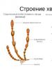
Scheme of the structure of the lungs: 1- trachea; 2 - bronchus; 3 - blood vessel; 4 - central (hilar) zone of the lung; 5 - apex of the lung.
Each lung is covered by a membrane (pleura). At the root of the lung, the pleura passes to the inner wall of the chest cavity. The surface of the pleural sac, which contains the lung, almost touches the surface of the pleura lining the inside of the chest. Between them there is a slit-like space - the pleural cavity, where a small amount of fluid is located.
During inhalation, the intercostal muscles lift and spread the ribs to the sides, the lower end of the sternum moves forward. Diaphragm (main breathing muscle) at this moment it also contracts, causing its dome to become flatter and lower, moving the abdominal organs down, to the sides and forward. The pressure in the pleural cavity becomes negative, the lungs passively expand, and air is drawn through the trachea and bronchi into the pulmonary alveoli. This is how the first phase of breathing occurs - inhalation.
When you exhale, the intercostal muscles and diaphragm relax, the ribs descend, and the dome of the diaphragm rises. The lungs are compressed, and the air from them is forced out. After exhalation there is a short pause.
Here it is necessary to note the special role of the diaphragm not only as the main respiratory muscle, but also as a muscle that activates blood circulation. Contracting during inhalation, the diaphragm presses on the stomach, liver and other abdominal organs, as if squeezing venous blood out of them towards the heart. During exhalation, the diaphragm rises, intra-abdominal pressure decreases, and this increases the flow of arterial blood to the internal organs of the abdominal cavity. Thus, the respiratory movements of the diaphragm, performed 12-18 times per minute, produce a gentle massage of the abdominal organs, improving their blood circulation and facilitating the work of the heart.
An increase and decrease in intrathoracic pressure during the respiratory cycle directly affects the activity of organs located in the chest. Thus, the suction force of negative pressure in the pleural cavity develops during inspiration and facilitates the flow of blood from the superior and inferior vena cava and from the pulmonary vein to the heart. In addition, a decrease in intrathoracic pressure during inspiration contributes to a more significant expansion of the lumen of the coronary arteries of the heart during the period of its relaxation and rest (i.e., during diastole and pause), and therefore the nutrition of the heart muscle improves. From the above, it is clear that with shallow breathing, not only ventilation of the lungs deteriorates, but also working conditions and the functional state of the heart muscle.
When a person is at rest, the act of breathing mainly involves the peripheral areas of the lung. The central part, located at the root, is less extensible.
Lung tissue consists of tiny air-filled bubbles - alveoli, the walls of which are densely intertwined with blood vessels. Unlike many other organs, the lungs have a double blood supply: a system of blood vessels that provide the specific function of the lungs - gas exchange, and special arteries that feed the lung tissue itself, the bronchi and the wall of the pulmonary artery.
Capillaries of pulmonary alveoli are a very dense network with a distance between individual loops of several micrometers (µm). This distance increases as the walls of the alveoli stretch during inspiration. The total internal surface of all capillaries located in the lungs reaches approximately 70 m2. At one time, up to 140 ml of blood can be in the pulmonary capillaries; during physical work, the amount of blood flowing can reach 30 liters per minute.
The blood supply to different parts of the lung depends on their functional state: the blood flow is carried out mainly through the capillaries of the ventilated alveoli, while in the parts of the lungs that are turned off from ventilation, the blood flow is sharply reduced. Such areas of lung tissue become defenseless when pathogenic microbes invade. This is what in some cases explains the localization of inflammatory processes in bronchopneumonia.
Normally functioning pulmonary alveoli contain special cells called alveolar macrophages. They protect lung tissue from organic and mineral dust contained in the inhaled air, neutralize microbes and viruses and neutralize harmful substances (toxins) released by them. These cells pass into the pulmonary alveoli from the blood. Their lifespan is determined by the amount of inhaled dust and bacteria: the more polluted the inhaled air, the faster the macrophages die.
From the ability of these cells to phagocytose, i.e. to the absorption and digestion of pathogenic bacteria, the level of general nonspecific resistance of the body to infection largely depends. In addition, macrophages clear the lung tissue of its own dead cells. It is known that macrophages quickly “recognize” damaged cells and move towards them to eliminate them.
The reserves of the external respiration apparatus, which provides ventilation to the lungs, are very large. For example, at rest, a healthy adult takes an average of 16 inhalations and exhalations per minute, and in one breath approximately 0.5 liters of air enters the lungs (this volume is called tidal volume); in 1 minute this will amount to 8 liters of air. With a maximum voluntary increase in breathing, the frequency of inhalation and exhalation can increase to 50-60 per minute, the tidal volume - up to 2 liters, and the minute volume of breathing - up to 100-200 liters.
The reserves of lung volumes are also quite significant. So, in people leading a sedentary lifestyle, the vital capacity of the lungs (i.e., the maximum volume of air that can be exhaled after maximum inhalation) is 3000-5000 ml; during physical training, for example in some athletes, it increases to 7000 ml or more.
The human body only partially uses oxygen from atmospheric air. As you know, inhaled air contains on average 21%, and exhaled air contains 15-17% oxygen. At rest, the body consumes 200-300 cm 3 of oxygen.
The transition of oxygen into the blood and carbon dioxide from the blood into the lungs occurs due to the difference between the partial pressure of these gases in the air in the lungs and their tension in the blood. Since the partial pressure of oxygen in the alveolar air is on average 100 mm Hg. Art., in the blood flowing to the lungs, the oxygen pressure is 37-40 mm Hg. Art., it passes from the alveolar air into the blood. The pressure of carbon dioxide in the blood passing through the lungs decreases from 46 to 40 mm Hg. Art. due to its passage into the alveolar air.
The blood is saturated with gases that are in a chemically bound state. Oxygen is carried by red blood cells, in which it enters into a weak connection with hemoglobin - oxyhemoglobin. This is very beneficial for the body, since if oxygen were simply dissolved in the plasma and not combined with the hemoglobin of red blood cells, then in order to ensure normal tissue respiration, the heart would have to beat 40 times faster than it does now.
In the blood of an adult healthy person contains only about 600 g of hemoglobin, so the amount of oxygen bound to hemoglobin is relatively small, approximately 800-1200 ml. It can satisfy the body's need for oxygen only for 3-4 minutes.
Since cells use oxygen very energetically, its tension in the protoplasm is very low. In connection with this, it must continuously enter the cells. The amount of oxygen absorbed by cells varies under different conditions. It increases with physical activity. The intensely formed carbon dioxide and lactic acid reduce the ability of hemoglobin to retain oxygen and thereby facilitate its release and use by tissues.
If the respiratory center, located in the medulla oblongata, is absolutely necessary for the implementation of respiratory movements (after its damage, breathing stops and death occurs), then the remaining parts of the brain provide regulation of the finest adaptive changes in respiratory movements to the conditions of the external and internal environment of the body and are not vital necessary.
The respiratory center reacts sensitively to the gas composition of the blood: excess oxygen and lack of carbon dioxide inhibit, and lack of oxygen, especially with excess carbon dioxide, excites the respiratory center. During physical work, muscles increase oxygen consumption and accumulate carbon dioxide, and the respiratory center responds to this by increasing respiratory movements. Even a slight holding of breath (breathing pause) has a stimulating effect on the respiratory center. During sleep, with a decrease in physical activity, breathing is weakened. These are examples of involuntary regulation of breathing.
The influence of the cerebral cortex on respiratory movements is expressed in the ability to voluntarily hold the breath, change its rhythm and depth. Impulses emanating from the respiratory center, in turn, affect the tone of the cerebral cortex. Physiologists have found that inhalation and exhalation have opposite effects on the functional state of the cerebral cortex and, through it, on voluntary muscles. Inhalation causes a slight shift towards excitation, and exhalation causes a shift towards inhibition, i.e. inhalation is a stimulating factor, exhalation is a calming factor. With equal duration of inhalation and exhalation, these influences generally neutralize each other. An extended inhalation with a pause at the height of inhalation with a shortened exhalation is observed in people who are in an alert state with high performance. This type of breathing can be called mobilizing. And vice versa: an energetic but short inhalation with a slightly stretched, extended exhalation and holding the breath after exhalation has a calming effect and helps to relax the muscles.
The therapeutic effect of breathing exercises is based on improving the voluntary regulation of breathing. In the process of repeated breathing exercises, the habit of physiologically correct breathing is developed, uniform ventilation of the lungs occurs, and congestion in the pulmonary circle and in the lung tissue is eliminated. At the same time, other indicators of respiratory function improve, as well as cardiac activity and blood circulation of the abdominal organs, mainly the liver, stomach and pancreas. In addition, the ability to use different types of breathing appears to improve performance and for proper rest.
Respiration is the process of exchange of gases such as oxygen and carbon between the internal environment of a person and the outside world. Human breathing is a complexly regulated act of joint work of nerves and muscles. Their coordinated work ensures inhalation - the entry of oxygen into the body, and exhalation - the release of carbon dioxide into the environment.
The respiratory apparatus has a complex structure and includes: organs of the human respiratory system, muscles responsible for the acts of inhalation and exhalation, nerves regulating the entire process of air exchange, as well as blood vessels.
Vessels are of particular importance for breathing. Blood through the veins enters the lung tissue, where gases are exchanged: oxygen enters and carbon dioxide leaves. The return of oxygenated blood is carried out through the arteries, which transport it to the organs. Without the process of tissue oxygenation, breathing would have no meaning.

Respiratory function is assessed by pulmonologists. The important indicators are:
- Width of the bronchial lumen.
- Breath volume.
- Reserve volumes of inhalation and exhalation.
A change in at least one of these indicators leads to a deterioration in health and is an important signal for additional diagnosis and treatment.
In addition, there are secondary functions that breathing performs. This:
- Local regulation of the breathing process, which ensures the adaptation of blood vessels to ventilation.
- Synthesis of various biologically active substances that constrict and dilate blood vessels as needed.
- Filtration, which is responsible for the resorption and disintegration of foreign particles, and even blood clots in small vessels.
- Deposition of cells of the lymphatic and hematopoietic systems.
Stages of the breathing process
Thanks to nature, which came up with such a unique structure and function of the respiratory organs, it is possible to carry out such a process as air exchange. Physiologically, it has several stages, which, in turn, are regulated by the central nervous system, and only because of this they work like a clock.

So, as a result of many years of research, scientists have identified the following stages that collectively organize breathing. This:
- External respiration is the delivery of air from the external environment to the alveoli. All organs of the human respiratory system take an active part in this.
- Delivery of oxygen to organs and tissues through diffusion; as a result of this physical process, tissue oxygenation occurs.
- Respiration of cells and tissues. In other words, the oxidation of organic substances in cells with the release of energy and carbon dioxide. It is easy to understand that without oxygen, oxidation is impossible.
The importance of breathing for humans
Knowing the structure and functions of the human respiratory system, it is difficult to overestimate the importance of such a process as breathing.

In addition, thanks to it, gases are exchanged between the internal and external environment of the human body. The respiratory system is involved:
- In thermoregulation, that is, it cools the body at elevated air temperatures.
- Functions as the release of random foreign substances such as dust, microorganisms and mineral salts or ions.
- In the creation of speech sounds, which is extremely important for the social sphere of a person.
- In the sense of smell.
The body needs energy to function. We get it from food, but for the effective breakdown of nutrients (oxidation) with the release of energy, the presence of oxygen is necessary. This occurs in the mitochondria of cells and is called cellular respiration. Oxygen must reach every cell of our body, so its transport is carried out by two systems: respiratory and cardiovascular. In the process of respiration and oxidation of organic substances, carbon dioxide is formed. Its removal is also the work of these two systems. Gases easily penetrate cell membranes. The cessation of metabolism means the death of the body. All cells of our body, without exception, must be continuously supplied with oxygen. Molecules of fats, carbohydrates and proteins located inside the body, when they combine with oxygen, oxidize, as if they burn. As a result of oxidation, these molecules decompose, the energy contained in them is released, carbon dioxide and water are formed.
Oxygen begins its journey through the airways respiratory system together with inhaled air, the oxygen content of which is 21%. First it enters the nasal cavity. There is a system of winding passages in which the air is warmed, moistened, and purified. The warmed air passes into the nasopharynx, and from there into the oral part and into.
From above, the entrance to the larynx is closed by one of the cartilages - the epiglottis, which prevents food from entering the windpipe. In terms of its internal structure, the larynx resembles an hourglass: it consists of two small cavities communicating through a narrow glottis, which in a calm state is triangular in shape and quite large. The larynx passes into the trachea - a tube 11–12 cm long, consisting of cartilaginous half-rings, which gives it rigidity and promotes the free passage of air. At the bottom, the trachea is divided into two, entering the right and left lungs. The mucous membrane of the inner wall of the trachea and bronchi is covered with ciliated epithelium. Here the saturation of the inhaled air with water vapor and its purification continues. The bronchi, entering the lungs, continue to branch into smaller and smaller branches, which end in the smallest. These are bronchioles, at the ends of which there are alveoli filled with air. The pulmonary vesicles are braided from the outside by a dense network of capillaries and are so closely adjacent to each other that the capillaries are sandwiched between them. The walls of the capillaries and bubbles are so thin that the distance between air and blood does not exceed 0.001 mm.

Gas exchange occurs due to the diffusion of gases through the thin walls of the alveoli and capillaries.
Molecules of any gas, if their concentration is high, tend to penetrate through shells that are permeable to them to places where there are few of them.
The change between inhalation and exhalation is regulated by the respiratory center, which is located in the medulla oblongata. It is sensitive to the carbon dioxide content in the blood and does not respond to the oxygen content. From the respiratory center, nerve impulses go to the muscles that produce breathing movements.