Subchondral sclerosis of articular surfaces and endplates. Osteophytes Sclerosis of the articular surfaces of the intervertebral joints
Vertebral growths are formed during the ossification of ligaments and other tissues. In patients who are under the supervision of doctors, it is often noted that first a strong growth of osteophytes, after a temporary slowdown in growth and a new start.
Some doctors consider these growths to be a kind of protection against joint pathology. Osteophytes warn the joint against excessive movement so that it cannot collapse further. Spondylosis is diagnosed mainly in people over the age of 50, but the disease can occur at an early age. A person with a perfectly healthy spine has space between the vertebrae.
More about ostephytes.
In the natural state, these niches should not be filled with any substance. But in the event of a hernia, disc dystrophy, osteophytes, these spaces no longer serve the normal functioning of the ridge. The localization of the growths is not the same in different patients.
They can be located in the front and back. Most of them resemble hooks in shape. Sometimes they grow so that they almost touch each other at the folds. It is better to treat spondylosis with therapeutic methods in the initial stage.
The doctor prescribes a number of anti-inflammatory drugs. They eliminate swelling
It is important to drink a complex containing B vitamins, phosphorus, magnesium and calcium. A number of ointments, gels and creams are recommended for external use.
Quite effective, especially at the initial stages of the development of osteophytes, methods of physiotherapy exercises, massage, physiotherapy. Diagnostics is carried out by a specialist - an osteopath or orthopedist. The disc height is reduced. This is fraught with:
- dislocations;
- inflammation.
The disease develops in several stages:
- Increased bone tissue volume.
- Ossification of the disc or ligaments.
The disease also occurs when the cartilage cover is disturbed. Because of this, irritation of the periosteum occurs, the tissues begin to grow.

There are a number of factors that contribute to the onset of spondylosis:
- Hard physical work.
- Excess weight.
- Arthrosis.
- Endocrine system ailments.
- Sedentary lifestyle.
- Flat feet.
- Genetic predisposition.
- Diseases of the nervous system.
- Violation of metabolic processes.
The listed factors appear separately or simultaneously.
You should pay close attention to healthy eating.
Types of osteophytes
- Bone compact;
- Bony spongy;
- Bone-cartilaginous;
- Metaplastic.
Bony compact osteophytesBony spongy osteophytesBone-cartilaginous osteophytesMetaplastic fracture infections
- Traumatic osteophytes. They are formed as a result of various traumatic injuries of bones, for example, after fractures, cracks, etc. Most often, traumatic osteophytes are formed in the area of fusion of two bone fragments displaced as a result of a fracture. The addition of an inflammatory process in the fracture area increases the risk of bone outgrowth formation. The shape, configuration and location of these osteophytes can be varied, but most often they are localized in the area of the knee and elbow joints. In more rare cases, traumatic osteophytes are formed without a bone fracture in the area of periosteum detachment, ligament tears or ruptures of the articular bags;
- Degenerative-dystrophic osteophytes. Formed against the background of chronic, long-term inflammatory or degenerative diseases of the joints, such as, for example, arthrosis, osteoarthritis, spondylosis, etc. outgrowths begin to form. These outgrowths provide an increase in the area of the cartilage, which reduces the load on the joint. After a while, these cartilaginous outgrowths begin to ossify and beak-shaped osteophytes are formed. The appearance of such osteophytes is a sign of deforming arthrosis. The outgrowths severely restrict movement in the affected joint. Degenerative-dystrophic osteophytes usually form in large joints or on the vertebrae;
- Inflammatory osteophytes. They are formed against the background of infectious and inflammatory lesions of the bones, for example, with osteomyelitis, tuberculosis, brucellosis, rheumatoid arthritis, etc. As a result of the formation of pus, the bone melts, defects in the form of holes, dips, etc. of these defects, an active process of bone regeneration begins, which ends with the formation of osteophytes of various shapes and sizes. As a rule, inflammatory osteophytes are multiple, randomly located on the surface of the affected bone, including in the joint cavity;
- Tumor massive osteophytes. Formed in malignant tumors or metastases in the bones (for example, with osteosarcoma, Ewing's tumor, metastases of prostate or breast cancer, etc.). A tumor or metastases damage the bone, and active regeneration begins in this area, which leads to the formation of large osteophytes in the form of spurs or a visor. Osteophytes in bone tumors are formed on the affected bone elements, and in metastases of prostate or breast cancer - mainly on the vertebrae or the iliac crest;
- Endocrine osteophytes. Formed against the background of systemic changes in the structure of bones and skeleton due to endocrine diseases. For example, with acromegaly, all the outer surfaces of the bones are covered with osteophytes, and with diabetes mellitus, outgrowths are formed in the area of the phalanges of the fingers, etc.;
- Neurogenic osteophytes. Formed as a result of a violation of the nervous regulation of metabolic processes and bone growth against the background of neurological diseases, such as, etc.;
- Osteophytes of increased physical activity. They can form on the surface of the bones due to damage to the periosteum by sharp contractions of the muscles attached to it, or in the joints due to tears or pinching of the joint capsule during movement. Due to constant damage to the surface of the bone, the repair process starts in it, which does not slow down, does not stop in time due to frequent signals of new damage. As a result, osteophytes are formed. Usually, such osteophytes are formed in athletes or in people engaged in hard physical labor.
Treatment
It is quite possible to cure a patient in case of the formation of osteophytes in the early stages. To do this, use physiotherapy exercises, physiotherapy, medications, folk recipes. Surgical treatment is rarely used.
Physiotherapy
The goal of exercise therapy in this case is:
- Improvement of blood circulation and metabolic processes in the affected segment and the spine as a whole. This leads to a slowdown in the growth of the osteophyte and promotes the resorption of excess bone tissue.
- Strengthening the muscular corset and ligamentous apparatus. Good muscle tone in the back and strong ligaments help keep the spine in a physiological position and relieve unnecessary stress on the vertebrae and intervertebral discs.
The set of exercises is selected individually, taking into account the localization of osteophytes and the physical condition of the patient. A good effect is given by classes in water, especially with severe pathology of the spine, as well as in the elderly.
Physiotherapy
Procedures are used to help improve blood circulation, relieve existing puffiness and enhance tissue regeneration processes. The most commonly prescribed are magnetotherapy, laser therapy, ozokerite, mud therapy, paraffin therapy. According to indications, hirudotherapy, therapeutic baths, massage can be used.
Traditional medicine methods
After consulting with your doctor, you can use folk remedies. For many years, the following recipes have been used by the people for pain and salt deposits:
- Compresses with honey at night. It is believed that honey is able to draw out excess salts through the skin.
- Herbal tea on birch leaves, lingonberries, pine buds.
- Healing baths (coniferous, chamomile, chestnut). Baths take a course of 10 procedures with an interval of 2-3 days.
- Alcohol rubbing on lilac flowers and a golden mustache.
Medications
Medicines help relieve pain and muscle spasm during an exacerbation, but they do not relieve osteophytes. The following drugs are used:
- Nise,
- Ibuprofen,
- Nurofen,
- Voltaren,
- Ortofen,
- Ketanov.
Surgery
Surgical removal of osteophytes is rarely used. The indications for surgery are bone growths that significantly squeeze the nerve or vascular bundles, causing persistent pain syndrome and disruption of the internal organs.
Bone-cartilaginous osteophytes
This type occurs when the structure of the cartilage tissue changes. In a healthy joint, all surfaces are covered with a cartilaginous layer. It performs very important functions: thanks to the cartilage, the sliding of the articular elements relative to each other during movement is ensured, and not friction, which would otherwise destroy bone tissue. In addition, cartilage serves as a shock absorber.
But if a disproportionate load is regularly given to the cartilage tissue, if an inflammatory process occurs in the joints and their degenerative changes occur, the cartilage loses its density and elasticity. It dries up and begins to deform.
Then the bone tissue, the mechanical effect on which increases, begins to grow. The formation of osteophytes in this case is a protective reaction of the body - thus it tries to increase the area of the joint and distribute the load. In this case, they often develop.
The site of localization of osteochondral osteophytes is large joints, knee or hip.
Osteophyte treatment
By itself, the identification of osteophytes is not enough to initiate treatment. It is imperative to establish the reason for their appearance. It is believed that if the growths do not cause pain and do not reduce mobility, then their treatment is not necessary.
If there is a severe pain syndrome due to nerve entrapment, then it is necessary to remove them surgically. An operation is never performed just to eliminate osteophytosis. First of all, the main problem in the joints and bones is eliminated. What type of surgery will take place and to what extent depends on the degree of joint damage.
For example: osteophytosis of the knee joint was diagnosed, treatment with conservative methods, as well as treatment with folk remedies, did not bring results, an operation is indicated. In this case, first it is necessary to carry out the correct alignment of the elements of the knee joint, if necessary, remove the destroyed parts of the bones and cartilage. If required, completely worn out cartilage is removed and replaced with mosaic grafts, and damaged bones are replaced with titanium implants.
Thus, osteophytosis is a consequence of other pathologies or injuries in a fairly advanced form. Its treatment is only a stage in the complex therapy of the main disease.
How to get rid of osteophytes
How to get rid of osteophytes in the spine, and can osteophytes dissolve? Doctors hear these questions every day.
At the initial stages of the formation of the pathological process, experts recommend to their patients conservative treatment of bone spines of the spinal column, which is a symptomatic therapy of pain syndrome and prevents the progression of the disease.
In order to relieve pain, patients are prescribed analgesics and non-steroidal anti-inflammatory drugs. To relieve the pathological tone from skeletal muscles, the patient is prescribed muscle relaxants and B vitamins.
At the initial stages of the formation of osteophytes, the doctor prescribes drug treatment
The peculiarities of the therapy of a pathological condition largely depends on the localization of the bone formations. So, the treatment of osteophytes of the cervical spine should include additional local use of ointments and gels with anti-inflammatory action, which will eliminate local edema and restore normal blood flow to the brain.
Before getting rid of osteophytes in the cervical spine, it is necessary to make a detailed examination of the affected area in order to find out the true nature of the origin of the spines, their size and placement in relation to the large vessels of the neck.
Treatment of osteophytes of the lumbar spine, as well as treatment of osteophytes of the thoracic spine, should be complex with the use of medications and that improve the trophism of cartilage tissue.
What exercise therapy exercises can be performed - see the video: https: //www.youtube.com/embed/LrURE7cftZw
Good results can be achieved if you use a massage bed and special orthopedic mattresses for osteophytes. Studies show that such techniques can significantly reduce the size of the thorns, stop the progression of the pathological process and prevent the development of new bone formations.
Surgical treatment of spinal osteophytes is indicated for patients who have:
- diagnosed with spike neoplasms of especially large sizes, which limit human mobility;
- there is no positive therapeutic effect from drug and physiotherapy treatment;
- removal of osteophytes in the cervical spine will allow the normal blood supply to the brain to be resumed and prevent the development of severe forms of ischemia in the head of the central nervous system in the patient.
Surgery has two goals:
- elimination of compression of the nerve roots;
- the actual removal of ossified spines with a part of the bone, which is then replaced by a prosthesis.
Before removing the osteophytes on the spine by surgery, the doctor carefully examines the patient's medical history and draws conclusions about the presence or absence of contraindications to surgery.
Diagnosis and treatment of the disease
The presence of osteophytes is diagnosed with X-ray, CT, or MRI. In some cases, for example, to study the state of adjacent tissues, several methods are required. The very fact of detecting bone growths is not of particular value for a specialist, but it is evidence of some kind of violation. It is at its identification that the further actions of the doctor are directed.
Osteophytes that do not cause discomfort cannot be treated. The main therapy will be aimed at eliminating the pathology that led to their development. If the outgrowths cause pain, injure the surrounding tissues, lead to a decrease or loss of working capacity, then I use conservative or surgical treatment.
Drug treatment
It will not be possible to eliminate osteophytes completely or reduce them in size with the help of drugs. Medicines are designed to get rid of pain, prevent inflammation in neighboring tissues, and normalize metabolism. For osteophytosis, the following groups of drugs are used.
- NSAIDs (diclofenac, indomethacin, ibuprofen).
- Corticosteroids (hydrocortisone).
- Vitamin complexes with mineral components (neurodiclovite, magnerot).
- Chondroprotectors (chondroxide).
Medicines are prescribed in the form of ointments and gels, injections or tablets (capsules).
Physiotherapy methods
As with drug therapy, the physical methods of influencing osteophytes are the same for all species (except those located on the vertebrae). Physiotherapy helps to eliminate symptoms, restores metabolism in tissues, and prevents further growth of formations. The earlier treatment is started, the greater the effect can be achieved.
With ostephytosis, appoint:
- electrophoresis;
- ultrasound;
- diadynamic therapy;
- acupuncture;
- magnetotherapy;
- vibration impact;
- laser treatment;
- shock wave therapy.
Shock wave therapy deserves special attention. It not only relieves symptoms, but also promotes the softening and destruction of osteophytes. In uncomplicated cases, this technique leads to the complete disappearance of bone growths.
ATTENTION! Shock wave therapy is not performed with osteophytes located along the spinal column due to the close location of the nerve roots. ... Physiotherapy and massage
Physiotherapy and massage
Massage procedures for osteophytes are used only on adjacent areas (the outgrowths themselves are not massaged!). Regular massage helps to prevent stagnation in muscle and connective tissue, improve blood circulation, eliminate metabolic products and other positive changes.
A set of therapeutic exercises is aimed at achieving the same goals. In difficult cases, it is developed by a doctor and performed in conjunction with an instructor. With not very pronounced osteophytosis, gymnastic exercises of exercise therapy can be found independently on the Internet. However, they must be performed, having previously agreed on the intensity and duration of the sessions with the attending physician.
Orthopedic aids
The use of special devices makes it possible to relieve the part of the body on which osteophytes have appeared. These can be: orthoses, insoles, corsets, tapes and others. In most cases, such mechanisms are used for osteophytes of the foot (heel spur or phalanges). They evenly distribute the load on healthy tissues, preventing pressure on the diseased area. Sticky tapes - tapes - relieve the joints of the limbs, spine.
Surgical intervention
With the massive development of osteophytes, they become the cause of disability. Persistent pain, symptoms corresponding to the location of the outgrowths (for example, immobilization, compression of the nerve roots), which are not eliminated with drugs and physiotherapy, are an indication for surgery. Surgical intervention is carried out by cutting off the bone growth. If necessary, a joint endoprosthesis is installed. The duration of the recovery period depends on the degree of damage to the bone and connective tissue, adjacent structures, the complexity of the operation, and is at least 2 months.
Types of osteophytes
There are several types of osteophytes:
- post-traumatic;
- degenerative-dystrophic;
- massive;
- periosteal;
- osteophytes resulting from systemic changes in the skeleton;
- neurogenic origin.
Post-traumatic osteophytes are the result of various damage to bone structures.
The appearance of such growths is possible with the preservation of the bone itself with a tear in the periosteum, which hardens over time, turning into an osteophyte.
Most often, this type of growths appears with dislocations of the elbow and knee joints, accompanied by the separation of the ligaments and rupture of the bursa. In the spine, post-traumatic osteophytes are rare.
Degenerative-dystrophic bone growth is manifested in such a disease as deforming arthrosis.
In this case, there is a slight limitation of joint mobility, without bone degradation.
The exception is cases of deforming spondylosis, as a result of which the surfaces of the joint are fused and its mobility is completely lost.
Such growths are subdivided into:
- general osteophytes - occur with senile arthrosis;
- local nature - are the result of overloading the local joint. In this case, the elasticity of the cartilage is lost and growths in the form of a beak are formed on the bone, which cover the joint, limiting its movement. In rare cases, the mobility of certain parts of the vertebra is lost.
Massive, or so-called marginal, osteophytes develop when:
- malignant bone tumors;
- metastases of breast or prostate cancer.
On an X-ray, they are seen in the form of a spur or visor, which is one of the important signs during the diagnosis of the disease.
Due to a violation of the growth process of cartilage, osteophytes can appear in benign tumors.
After inflammatory processes, the growth of periosteal osteophytes can be observed, which are formed from the beneficial components of the periosteum.
As a result of endocrine disorders and due to systemic changes that occur for this reason in the skeleton, osteophytes can also appear.
Hypertrophy of the bone relief leads to the formation of growths on:
- sciatic tubercle;
- nail phalanges;
- thigh skewers, etc.
The appearance of osteophytes can also provoke psychological disturbances - for example, the formation of growths during disorderly bone formation can be observed during a nervous breakdown.

Also, osteophytes are classified according to the place of their localization:
- anterior - appear on the anterior parts of the vertebral bodies. Formed mainly in the thoracic region and rarely cause pain;
- posterior - "grow" on the posterior surfaces of the spine. Unlike the anterior ones, their formation is accompanied by a strong pain syndrome, since there is mechanical pressure on the nerve trunks of the intervertebral foramen;
- the anterolateral bony outgrowths have a horizontal direction and an unusual shape in the form of a bird's beak. Sometimes there are so-called kissing osteophoritis, in which the ends are pointed and approach each other. Formed in areas with the greatest pressure, where there is a change in the intervertebral discs;
- posterolateral arises mainly in the cervical vertebra and cause compression of the spinal cord.
Modern approaches to treatment
Treatment of osteophytes of the knee joint in practice is implemented through the use of conservative methods of therapy, physiotherapy, recipes of traditional medicine, and in especially difficult cases - surgical correction. Medical treatment of thorns has several goals:
- relief of pain syndrome;
- reducing the manifestation of a local inflammatory reaction;
- improving the trophism of damaged tissues;
- restoration of damaged areas of cartilage and a normal amount of intra-articular fluid.
Before treating osteophytes of the knee joint, the doctor necessarily conducts a detailed examination of the patient and diagnoses concomitant diseases that may be a contraindication to taking medications.
Before prescribing treatment, the doctor will carefully study all the features of the course of the disease.
As a therapy for osteophytes, the patient can be prescribed non-steroidal anti-inflammatory drugs, analgesics, chondroprotectors, hyaluronic acid, as well as drugs to improve metabolic processes and blood flow in tissues (medical bile, vitamin complexes).
How to remove osteophytes in the knee joint using physiotherapy?
For this purpose, the patient is offered to undergo several courses of galvanization, electrophoresis, phonophoresis with hormones and analgesics, ultrasound therapy, take baths with turpentine, and massage on the damaged joint.
Shockwave therapy devices loosen the osteophytes, and then the osteophytes dissolve altogether. How the session of radiation by the method of shock wave therapy takes place - see the video: https: //www.youtube.com/embed/pvVhAXzd9F0
How to get rid of osteophytes in the knee joint when all conservative methods were used, but did not have the desired therapeutic effect?
In this case, specialists offer patients with advanced forms of the disease and large size of bone formations, which prevent a person from moving normally, surgical treatment, which involves excision of growths with a part of the bone and its subsequent plastic surgery.
As a rule, in case of small osteophytes, patients are recommended a procedure with minimal access and traumatism to prevent the development of postoperative complications.
Causes of osteophytes
Age-related changes in the structure of the spine are considered to be the main causes of osteophytes. Indeed, over the years, a person forms various deposits or other modifications in bone tissues, which require hospital treatment.
Also, the correct posture of a person plays a huge role. Indeed, when sitting at a table for a long time in an unnatural posture or heavy physical exertion while walking leads to stoop and the appearance of osteophytes. But in addition to the main reasons for the growth of bone tissue, other common factors should be noted:
- Spine diseases (osteochondrosis, scoliosis, kyphosis). As a result of displacement or deformation of the spine, friction of neighboring vertebrae occurs, which becomes the result of the appearance of degenerative-dystrophic osteophytes;
- Hereditary predisposition;
- Disrupted metabolism;
- Inflammatory process. As a result of inflammation of one or more vertebrae, periosteal osteophytes appear
- Injuries. Post-traumatic osteophytes occur at the sites of vertebral fractures or joints of the spine;
- Flat feet;
- Oncological diseases. The emergence of a malignant tumor provokes the occurrence of metastases in the spine, thereby forming massive osteophytes;
- Constant physical activity;
- Excess weight.
//www.youtube.com/embed/bMpwxV2wiVw
Types of growths
The term "osteophyte" means a certain type of bone growth, which is provoked by a specific cause. Osteophytes differ in their location, structure and causative factor.
According to the cellular structure, there are such types of osteophytes:
- spongy;
- metaplastic;
- compact;
- cartilaginous.
Spongy is formed from the spongy substance that forms the articular surfaces. Also small bones are made of it. For example, vertebrae, wrists, ribs, etc. Typically, spongy osteophytes appear due to heavy stress on the bones.
Metal osteophytes appear when the cellular composition of bones is disturbed. Usually formed after injuries, fractures and other damage to the bone. In some cases, it may appear against the background of inflammatory processes or infections.
 As for compact osteophytes, they are formed from the outer bone layer. The latter is well developed in the tibia, femur, ulna, and radius, for this reason compact osteophytes appear on these bones. They are usually found on the feet and toes.
Cartilaginous osteophytes are found in the joint cavity, where the surface is covered with cartilage. When the latter is exposed to stress, it begins to thin, the bone grows, and osteophytes are formed.
Types of osteophytes that appear for a specific reason:
- Degenerative-dystrophic osteophytes. They appear as a result of long-term inflammatory diseases of the joints (arthrosis, spondylosis).
- Tumor - appear in the presence of metastases or malignant tumors, for example, with sarcoma, cancer metastases, etc. The tumor damages the bone, as a result, regeneration begins, leading to the formation of osteophytes.
- Traumatic osteophytes - appear when bones are injured. Often found in the area of fusion of displaced bone fragments.
- Inflammatory. Appear in the presence of inflammatory lesions (osteomyelitis, brucellosis).
How to get rid of osteophytes
Comprehensive treatment
When osteophytes appear, this is forever. They do not dissolve by themselves. The task of doctors is to make life easier for the patient and slow down the development of bone growths. But otherwise, the treatment of osteophytes is carried out in the same way as for other diseases of the spine. Complex therapy has several components:
- Medicines. First, as soon as a problem arises due to bone growths, anti-inflammatory drugs are prescribed. But if the pain becomes severe, then pain relievers are additionally used. It can be Analgin, Butadion, etc. Or muscle relaxants are additionally prescribed. Warming ointments (Viprosal, etc.) help relieve pain.
- Physiotherapy. Along with drugs, special physical exercises are prescribed. But with inflammatory processes or severe pain, you cannot go to exercise therapy. In any case, the first exercises should be simple, and the load should be increased carefully and gradually.
- Massage. Manual therapy also successfully fights bone growth. Specialists in this field work according to certain methods. Massage improves blood flow to damaged vertebrae and joints. It helps relieve muscle spasms and partially or completely restores motor function. There is a gradual regeneration of cartilage tissue.
- Epidural injections with steroids. Injections are prescribed if inflammatory processes have begun in the joints, which are accompanied by edema. Then the patient is injected with steroids. The effect of the injection is only temporary.
- Physiotherapy. There are two techniques used in this area. HILT therapy is done using a laser that reaches deep-seated osteophytes. In shock wave therapy, acoustic waves are used.
Surgical intervention
If complex therapy does not help, then the disease is severe. In this case, surgical intervention is required. The doctor performs an operation during which the bone growths are removed. But if the nerve endings were compressed for a very long time, then after surgery, the patient may show neurological symptoms. This means that irreversible changes have already taken place in the fibers.
Treatment with folk remedies
Treatment of osteophytes with folk remedies has many ways to deal with bone growths. Or at least the recipes help relieve pain. These are mainly decoctions and herbal infusions.
- Elder. To prepare infusion from it, you will need 1 tbsp. l. berries, which are poured with a glass of boiling water. Then they are heated in a water bath for a quarter of an hour. The infusion is cooled and filtered. Then half a glass is taken 2 to 3 times a day.
- Hawthorn. For infusion, only flowers are taken from the plant. One handful is poured with three glasses of boiling water. Then it is infused for 30 minutes. After the infusion is cooled and taken in 3 tbsp. l. half an hour before meals.
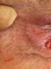
Why arises
The main factors in the appearance of bone growths on the back of the foot are degenerative and inflammatory processes in the tendons, which provoke the appearance of small cracks or tears. This is due to the uneven distribution of loads on the heel and surrounding tissues. If this kind of injury is only a single one, then it quickly passes. However, their large number provokes severe damage to the bone and adjacent tissues. As a result, the affected area becomes inflamed, and a small formation develops in its place. If there is no treatment, the bone continues to grow, causing pain and discomfort to the person. Heel osteophytes can develop under the influence of factors such as:
- overweight
- wearing uncomfortable shoes
- flat feet,
- infectious diseases,
- metabolic failure,
- damage to nerve endings.
In the plural, calcaneus masses are very painful, especially in the morning. Therefore, in order not to feel painful sensations, a person transfers the weight of his weight to the front of the leg, which provokes a change in gait.
Diagnostic methods
When a patient calls for help, a specialist conducts a neurological examination, during which he will be able to identify signs of compression of the spinal cord and roots.
Based on the history of the disease, the patient's complaints and the result of the examination, the doctor prescribes a further examination.
In cases where osteophytes are large, their detection is possible by simple palpation.
In this case, the specialist will be able to freely feel the hills in the form of thorns and tubercles in a certain area of the spine.

To confirm the diagnosis and at earlier stages of the disease, when it is not yet possible to probe the growths, the following diagnostic methods are used:
- radiography;
- Magnetic resonance imaging;
- CT scan.
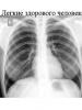
X-ray is a research method based on the use of X-rays.
It is completely painless and helps to identify the presence of bone formations. According to the results of X-ray, the degree of development of the pathology and the shape of the growths are determined, according to which the final diagnosis is made.
In the picture, osteophytes are bone formations of various sizes and character, localized along the edges of the vertebrae.
A more detailed description of changes in the bone and soft tissue structures of the spine can be obtained by magnetic resonance imaging and computed tomography.
Subchondral sclerosis of the articular surfaces is the process of death of healthy cells of the joints and their subsequent replacement with connective tissue. It is more of an X-ray term than an individual disease. Often, subchondral sclerosis becomes a sign of advanced osteoarthritis.
Reasons for the development of subchondral sclerosis
The subnerral ball is the layer of tissue that adjoins the cartilage. Unfortunately, with age, it can harden and grow. This is due to the fact that the blood supply to the subchondral layer is disrupted due to atherosclerosis or other vascular pathologies. Due to the violation of blood flow to the subchondral layer, the structure of the cartilage is disrupted. This is one of the pathogenetic mechanisms of osteochondrosis and the appearance of osteophytes.
The free process develops with age. The elasticity of the structures that surround the joint is lost, cartilage is destroyed and sclerosis is formed. Subchondral sclerosis and osteophyte manifestation is a diffuse phenomenon, most often spread over the spine. Large growths provoke pain during movement.
Subchondral sclerosis and osteosclerosis can develop for such reasons:
- degenerative processes in the joints (osteoarthritis);
- constant, uneven physical activity on the joints;
- excess weight;
- diabetes mellitus and atherosclerosis of the lower extremities;
- autoimmune pathologies.
 Subchondral sclerosis often affects the hip joints of patients over 50 years of age. This is a symmetrical process, accompanied by morning stiffness and pain during physical exertion.
Subchondral sclerosis of the joints of the lower extremities is accompanied by intermittent claudication, soreness at the end of the working day.
Subchondral sclerosis treatment
Unfortunately, subchondral sclerosis is untreated in 80% of cases. You can only stop the progression of pathology. It is not known until the end which of the factors influences the development of the disease and it is impossible to cure it with one pill.
But if you do not engage in treatment at all, this will lead to immobility of the limb or spine. Over time, ankylosis will develop against the background of subchondral sclerosis.
What drugs are prescribed for a patient with osteosclerosis:
- Pain relievers and anti-inflammatory drugs from the NSAID group (Diclofenac, Meloxicam, Celecoxib).
- Chondroprotectors. The funds have unproven effectiveness, therefore they are used only as an auxiliary method.
- B vitamins. Relevant for vitamin deficiency, polyneuropathies. Combined drugs are prescribed (Milgamma, Kombilipen).
With severe pain in the joints, the patient needs to do novocaine blockade. They are performed by a vertebrologist or neurologist. 
It is possible to connect a course of physiotherapeutic methods in the treatment of osteosclerosis:
- magnetic waves;
- phonophoresis and electrophoresis;
- paraffin and mud applications.
The extreme degree of joint immobility is stopped by a surgical method - endoprosthetics.
It is important to engage in dosed physical activity or exercise therapy.
Basic rules for a patient with osteosclerosis, which should be followed at home:
- monitor body weight by body mass index;
- do not injure bones and joints;
- engage in dosed physical activity;
- avoid infectious diseases and inflammation of the joints.
The unpleasant sensation of discomfort, accompanied by joint pain, is familiar to many people, especially the elderly. It happens that due to serious physical exertion, such manifestations are of a periodic, short-term nature. In such cases, you can cope with them yourself with the help of various pain relievers. But when joint problems develop into a serious illness, a person is forced to see a doctor.
Among the variety of diseases affecting the joints, such ailment as subchondral sclerosis of the articular surfaces deserves special attention. With this pathology, degenerative processes develop in the cartilage or bone tissue. As a result, the articular surface undergoes serious changes. By and large, subchondral sclerosis cannot be considered an independent disease. It is rather an X-ray symptom, indicating that the cause of this ailment is other pathologies of a degenerative-dystrophic nature occurring in the body. Therefore, it is rather difficult to get rid of subchondral sclerosis without treatment of the underlying disease.
In subchondral sclerosis, due to inflammation, trauma or aging of the body, the connective tissue of the joint is affected. As a result, bone density increases, and it grows. Such a pathological process leads to the fact that irregularities appear on the articular surface, bone outgrowths - osteophytes - are formed. The appearance of such irregularities increases friction, which is the cause of pain. With an incorrect diagnosis, untimely treatment, further growth of outgrowths can block the joint and completely immobilize it.
According to the observations of doctors, subchondral sclerosis most often develops against the background of such degenerative-dystrophic diseases as arthrosis and. Of course, the causes of their occurrence and symptoms are somewhat different, but arthrosis and osteochondrosis have one common feature: the development of these diseases, as a rule, always leads to the formation of osteophytes. Only with arthrosis, osteophytes are formed along the edges of the joints, and in the presence of osteochondrosis, bone outgrowths appear along the edge of the vertebrae.
Unfortunately, such formations, which characterize subchondral sclerosis, cannot be completely cured. With the help of drug therapy, you can only stop the further progression of the disease and prevent the formation of new bone growths. Moreover, doctors involved in the treatment of this pathology rarely resort to surgical intervention, since even an operation is not able to radically change the situation.
Distinguish between primary and secondary forms of development of subchondral sclerosis. In the primary form, pathological changes in the articular surface occur in a healthy joint. Pain syndrome occurs due to the load on the musculoskeletal system, and subsides during rest. In the secondary form of the disease, an already damaged joint is affected after arthritis or an injury. In most cases, subchondral sclerosis affects the ankle, knee, and hip joints. The most dangerous consequences of this pathology are the formation of bone spurs and complete immobility.
Doctors note that the treatment process for subchondral sclerosis takes a long time and requires a lot of effort. In the case of severe pain, the patient is prescribed non-steroidal anti-inflammatory drugs and analgesics. The patient needs to regularly perform special exercises to improve blood circulation in the affected joints and flexibility of movement. In addition to the main measures of influence, hardware physiotherapy, acupuncture helps, a semi-bed or orthopedic regime is possible.
One of the important conditions for the treatment of subchondral sclerosis of the articular surfaces is the limitation of physical activity. To reduce the likelihood of progression of the disease, it is necessary to monitor your own weight, since each extra kilogram significantly increases the load on the joint. Therefore, at the same time with therapeutic measures, a dietary diet should be observed.
It is well known that joint pain is an inevitable manifestation of the aging of the body. Timely visits to doctors, maintaining a healthy lifestyle will help to identify a dangerous disease at an early stage. You should not wait for the dangerous consequences that subchondral sclerosis often leads to. Take care of yourself!
Degenerative diseases of bones and joints of the musculoskeletal system occurs under the influence of many factors. Subchondral sclerosis develops mainly in the elderly, has an irreversible course and greatly complicates the patient's life.
What is subchondral osteosclerosis?
Subchondral osteosclerosis is a pathological condition in which compaction of bone tissue develops directly under the lower surface of the cartilage, disrupting the blood supply and structure of the latter. Joint sclerosis is dangerous because it leads to early disability and limited motor activity. Dense but thinned bone breaks with minor injuries, and sometimes under the weight of its own body.
Reasons for the formation of subchondral sclerosis
Bone disease does not develop suddenly, but is formed for many years under the influence of reasons that directly or indirectly affect the health of the musculoskeletal system. The factors provoking the development of subchondral osteosclerosis are divided into two groups.
Endogenous (internal) factors include:
- Physiological aging of the body. Disruption of mineral metabolism, a change in the balance between "old" and "new" bone cells and other signs characteristic of an elderly organism lead to osteosclerosis.
- Hereditary nature of development.
- Endocrine disorders such as diabetes mellitus, hyperparathyroidism.
- Metabolic disorders, such as Wilson-Konovalov disease, gout.
- Vascular diseases acquired during life and impairing blood circulation in the extremities.
- Autoimmune diseases in which the body's own immune cells damage the body. These include systemic lupus erythematosus and rheumatoid arthritis.
Exogenous (external) factors include:
- Musculoskeletal system injuries. With regard to the development of subchondral osteosclerosis, fractures of the articular surfaces are especially dangerous injuries.
- Microdamages arising in dancers, athletes, military men under the influence of prolonged and excessive loads on the feet and knees.
- Overweight due to improper lifestyle and overeating is one of the most destructive factors for the musculoskeletal system. It promotes increased trauma and passive destruction of the skeleton.
- Restriction of motor activity, which contributes to the weakening of the auxiliary structures of the supporting apparatus, impaired outflow or inflow of intra-articular fluid.
Stages of development of osteosclerosis
The course of subchondral bone diseases is divided into 4 stages. The transition from the initial stage to the last stage is accompanied by characteristic radiological symptoms.
- The initial stage is characterized by marginal osteophytes that form on the surface of the joints.
- Moderate subchondral sclerosis corresponds to stage 2. The radiograph shows a narrowing of the inter-articular space. The focus of sclerosis under the joint is determined in the form of enlightenment (negative in the picture) against the background of relatively healthy bone tissue.
- At stage 3, the joint space is significantly narrowed, osteophytes increase in size, damage the cartilage due to friction of the deformed surfaces. Clinically, this is manifested by joint pain during movement and impaired mobility. Often, at this stage, a "articular mouse" appears - a fragment from an osteophyte or a deformed surface, which is chipped off under the influence of additional traumatic factors. Cartilage destruction is noticeable during arthroscopy.
- The fourth stage is characterized by significant joint deformities with the formation of flat, incongruent surfaces. The inter-articular gap is not determined, osteophytes wedge into the bone, provoking chips, which are determined in the periarticular space. In the epiphyses of the bone on X-ray, the alternation of extensive foci of osteosclerosis with areas of osteoporosis is noticeable. During arthroscopy, the cartilage is completely destroyed and cannot be visualized. A person loses the ability to move independently, feels constant pain, and it is also impossible to perform active and passive movements.
Forms of subchondral osteosclerosis
According to the prevalence of osteosclerosis in the human skeleton, the following clinical forms can be distinguished:
- The limited form looks like a focus of osteosclerosis against the background of healthy tissue within the same anatomical formation.
- Multiple sclerosis affects more than 1 limb or anatomical area. Diseases causing a common form include Paget's disease, Leri's melorheostosis, and malignant neoplasms with metastases.
- Systemic osteosclerosis occurs under the influence of many factors and totally affects the skeleton.
Subchondral sclerosis of the spine

The most problematic form of the disease is subchondral osteosclerosis of the endplates of the vertebral bodies. The development of sclerosis in one vertebra is often not felt by a person. However, when osteophytes gradually wedge in and put pressure on the nerves emanating from the spinal cord. Sclerosis of the endplates of the spine affects different parts of the bone structure, causing the following symptoms:
- Sclerosis of the endplates of the cervical vertebrae is the most insidious, as it disrupts important functions of the body. Compression of nerves and blood vessels leads to dizziness and ringing in the ears, vision decreases, deafness progresses, coordination of purposeful actions is impaired. Bad prognostic signs are - violation of the respiratory rhythm, increased heart rate and cardiac pain, decreased memory, attention. At the slightest movement of the neck, a dull or "shooting" pain appears. Subchondral sclerosis of the endplates of the cervical region leads to a decrease in sensitivity and muscle strength. Pronounced leads to a complete loss of movement in the hands due to compression and destruction of nerve fibers at the level of 4-7 vertebrae of the neck.
- Subchondral sclerosis of the endplates of the thoracic vertebral bodies is manifested by impaired breathing, a noticeable distortion of posture. Pain in this area hinders movement.
- Sclerosis of the lumbar spine is manifested by shooting pain when bending and turning the body. With the progression of the disease, weakness appears in the legs, due to which a person may lose the ability to move independently.
It is obvious that subchondarial sclerosis of the endplates of the vertebral bodies requires timely treatment, without which there is a rapid development of neurological symptoms with severe motor and sensory disorders.
Osteosclerosis of the joints of the upper limb
Subchondral osteosclerosis of the articular surfaces of the bones of the upper limb at the initial stage of the degenerative process is manifested by a crunch during flexion and extension of the arm, which is not accompanied by pain. After a short period of time, a person develops a feeling of a foreign body that interferes with normal movement in the elbow joint.
With a pronounced deformation of the articular surface, the arm does not unbend, and any attempt to straighten the arm is accompanied by severe pain syndrome.
Osteosclerosis of the joints of the lower limb
Subchondral sclerosis of the hip joint is the most unfavorable localization of the degenerative-dystrophic process in the elderly. The development of osteosclerosis in this localization significantly increases the risk of hip fracture. If there is a deformation on the side of the acetabular surfaces, then the patient has aching pain in the lumbar and pelvis. With the localization of the pathological focus in the femur, the sensation of pain arises from the outside of the anatomical region of the same name. At first, the disease resembles subchondral sclerosis of the spine, but later there are signs of impaired movement in the hip joint, which confirms the true localization of the pathology.
Osteosclerosis of the knee joint begins with the appearance of characteristic "clicks" when moving in full. Pain is often caused by loosening of the ligaments. The walking process becomes more complicated, it is almost impossible to bend the leg and the person starts to move on "straight" legs or limps. Sclerosis of the articular surfaces of the knee without treatment leads to a wheelchair.
Diagnostics of the subchondral osteosclerosis

In diseases of the musculoskeletal system, methods for visualizing the structure of the bone and joint are of particular importance. These include:
- Radiography. The simplest and most accessible method is widely used to diagnose subchondral sclerosis.
- Magnetic resonance imaging (MRI). Despite the general approval of this method in the study of the nervous system and parenchymal organs for the musculoskeletal system, the method is of less value. This is due to the fact that the visualization of soft tissues on MRI is better than hard tissues, respectively, for examining bones it is less informative.
- CT scan. For the study of the musculoskeletal system, the method is informative. On CT scan, the hard tissue of the bone and joint is clearly visible, it makes it possible to identify the area of subchondral osteosclerosis with almost no errors.
Laboratory studies and other methods are used to carry out differential diagnosis with other diseases or in the case of an unclear clinical case.
Treatment methods
Treatment of osteosclerosis requires an integrated approach, including:
- Modification of lifestyle and diet.
- Medication.
- Surgery.
- Remedial sports activities.
Lifestyle changes are recommended for people with a sedentary lifestyle. Exercising and working out your joints every day will help slow down degenerative diseases as well as reduce excess body weight. There are no special restrictions on the diet, but it is not recommended to consume large amounts of salt.
The basic principles of the treatment of osteosclerosis involve the use of drugs from the following groups:
- Non-steroidal anti-inflammatory drugs (Indomethacin, Diclofenac).
- Chondroprotectors (Chondroitin).
- Preparations containing chondratin and glucosamine, which are necessary for the regeneration of cartilage tissue.
Surgical treatment of osteosclerosis is used in the last stages of osteosclerosis, when the articular surfaces are already completely deformed. Surgical interventions involve the installation of titanium prostheses, which restore the lost functions.
Exercise therapy is used during the recovery period, after an exacerbation of the underlying disease or its complications. A specially developed program is used to perform a number of exercises aimed at the rehabilitation of joints and bones.
Subchondral sclerosis is a degenerative lesion of the cartilage covering the inner surfaces of the joints, in which normal functional tissue is replaced by connective tissue that is unable to perform the required functions. At the same time, the bone tissue of the joints also begins to thicken and grow, forming growths.
This pathological process does not stand out as a separate disease, but is one of the manifestations of osteoarthritis of the joints and osteochondrosis of the spinal column. It does not develop immediately, but as the underlying disease progresses, if the causative factors are not eliminated, and the treatment is incorrect. Elderly people are more susceptible to subchondral sclerosis, but recently it has also been noted in young people.
Stages of subchondral sclerosis
The development of the disease takes place in stages:
- Initial subchondral sclerosis - the growth of bone tissue occurs only along the edges of the joint.
- Moderate subchondral sclerosis - osteophytes are visible on the x-ray, the joint space is narrowed, and the articular part of the bone is characterized by a lighter color.
- Stage III subchondral sclerosis - there is a significant narrowing of the joint space, large bone growths, the motor activity of the joint is significantly impaired.
- Stage IV subchondral sclerosis - osteophytes are very large, the articular surfaces of the bones are significantly deformed, the inability of the joint to full extension and flexion is noted.
Subchondral sclerosis of the knee joint - what is it?
The knee joint is very often affected by subchondal sclerosis. he is constantly exposed to high loads. Risk factors for the development of pathological processes in this joint are:
- overweight;
- hormonal disruptions;
- professional harm.
Pathology is detected in patients with deforming osteoarthritis of the knee joints, manifested by such symptoms as pain during exercise and at rest, crunching during movement, difficulty in flexion-extension of the knee. In this case, cracking, thinning of the cartilaginous tissue occurs, and its loss of strength and elasticity. A common consequence of subchondral sclerosis of the knee joint is the development of varus or hallux valgus.
Subchondral sclerosis of the spine - what is it?
Subchondral sclerosis of the endplates of the vertebral bodies of the spine is more often observed in the cervical spine, less often in the thoracic and lumbar. In this case, patients complain of chronic pain in the corresponding affected area, neurological complications (numbness of the extremities, dizziness, etc.), spinal deformities are also possible.
The main danger of the pathology of this localization is the increased risk of spontaneous compression fractures, which can occur even with minimal physical exertion. In the most advanced cases, there is partial or complete paralysis.
Subchondral sclerosis of the hip joint

This localization of pathology almost always complicates the course of arthrosis of the hip joint. The main manifestations in this case are: chronic pain in the hip area (in motion and at rest), limitation of the range of motion in the joint, the development of lameness.
Subchondral sclerosis of the hip joint is dangerous due to the increased risk of fracture of the femoral neck and aseptic necrosis of its head. Therefore, when a pathological process is identified, one should immediately start to prevent possible severe consequences. If treatment is not started on time, limb functions can be completely lost.